40 how to label gel electrophoresis images
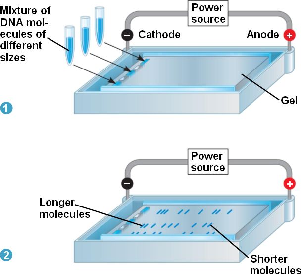
PDF 8/13/2009 Tutorial ImageJ Using ImageJ to Quantify Gel Images of interest go to Image/Crop to crop the selection.See below for screenshots. Enhancing the Gel Image This is a typical step when dealing with gel images. You need to adjust the histogram of the image. Please make sure not to blow-out (saturate) the whites. You want to make sure your image has enough dynamic range. Talk to me if you're confused. Gel Electrophoresis - an overview | ScienceDirect Topics For gel electrophoresis, a DNA sample is loaded at one end of a gel matrix (usually agarose or acrylamide) that provides a uniform pore size through which the DNA molecules can move. Application of a constant electric field causes DNA fragments (all have a uniform, strong negative charge) to migrate toward the cathode.
Annotating A Gel | Get Your Science On Wiki | Fandom Part 1. Photo Editing: 1.Take your JPG or PNG file of your Gel and open it with a photo editing program (GIMP). 2. Under "Image" --> "Transform" rotate your picture by 90 degrees so that your wells are on top of the page. 3. Using the Crop tool Cut out the black borders leaving only the gel. 4.

How to label gel electrophoresis images
A Complete Guide for Analysing and Interpreting Gel Electrophoresis Results let see some of the gel images of PCR fragments. 2% gel is required to separate PCR products because PCR products are the smaller fragments of DNA nearly ~100bp to ~1500bp. Image 1: The image is captured under the UV transilluminator instead of the gel doc system to show you the effect of EtBr on the gel electrophoresis results. PDF Lab 4: Gel Electrophoresis - Vanderbilt University This protocol uses a standard electrophoresis system. The agarose gel will be made by adding agarose powder (or tablets) to running buffer, boiling the mixture, then letting it cool into a gelatin-like slab. The agarose gel is run in a standard electrophoresis system, then visualized with a transilluminator. Pre-Lab Preparation Gel Electrophoresis: Definition, Principle, and Application Electrophoresis is a process used for the separation of macro and micro molecules in an electric field by applying charges at both the extents. The mixture of substances is spread in the supporting film. The supporting films are placed in a salt solution filled in a container, where one container holds a cathode and the other carries an anode.
How to label gel electrophoresis images. CHAPTER 10 Flashcards | Quizlet Please label the images to review the process of polymerase chain reaction and how its products can be analyzed using gel electrophoresis. Match the components of a typical PCR reaction with the function they serve. ... Please label the images to review the process of screening bacterial clones for those containing a donor gene. Other sets by ... Gel electrophoresis (article) - Khan Academy When a gel is stained with a DNA-binding dye and placed under UV light, the DNA fragments will glow, allowing us to see the DNA present at different locations along the length of the gel. The bp next to each number in the ladder indicates how many base pairs long the DNA fragment is. A well-defined "line" of DNA on a gel is called a band. Gel Electrophoresis - University of Utah Gel Electrophoresis. Have you ever wondered how scientists work with tiny molecules that they can't see? Here's your chance to try it yourself! Sort and measure DNA strands by running your own gel electrophoresis experiment. Click to unmute. See how gel electrophoresis is used in forensics. Can DNA Demand a Verdict? PDF Gel Electrophoresis Size Marker - dia-m.ru Labeling in four positions via the terminal EcoR I generated recessed ends is possible, especially with the DNA ladder 100 bp (A3470) and the DNA Ladder Mix 100 - 5000 (A3660). For the labeling of the DNA, the product is simply dissolved in TE buffer or bidistilled water. DNA staining with methylene blue
Analysis of protein gels (SDS-PAGE) - Rice University Calibrate the gel using standards of known molecular mass (set up a standard curve if necessary) Select polypeptide bands in the lane (s) of interest to be analyzed and identify them by some generic label (e.g., a, b, c,... or 1, 2, 3,...) Estimate molecular mass or relative molecular mass for each band of interest How to quantify each band in gel electrophoresis? - ResearchGate you can do an analytical curve in a 1d gel, with known amounts of bsa for example, use photoshop to quantify the pixels (the curve would be pixels x protein mass you applied for each well) and then... ImageJ for Editing & Labelling PCR Gel Image - YouTube This Tutorial is all about how to quickly Edit & Label PCR Gel Image Using ImageJ software. Presented by - Elvis SamuelJoin Our Telegram Channel for free Sof... GelAnalyzer GelAnalyzer 19.1 Analyze gel images from any source Use your digital camera, smartphone, or gel doc system to obtain images. GelAnalyzer will take care of the rest. Automatic lane and band detection With full manual control over adding, modifying, and deleting lanes and bands. Fix run distortions through Rf calibration
Solved Please label the images to review the process of - Chegg Science. Biology. Biology questions and answers. Please label the images to review the process of polymerase chain reaction and how its products can be analyzed using gel electrophoresis. Cycle 1 Priming MEGF1-44- MER Sagan 1o Bened d Haut 94C New strand Strands pere SOM 500 5 PERAN Original strands Cydia Amplicon Pos Am Ciprusside parcial ... PDF Gel Electrophoresis: How Does It Work - Purdue University a. After you find out what dyes you are using, draw a picture of the gel and the wells. Label which dyes you will put in each well. b. When you load a gel, it is very important that you do not damage the gel in any way. You must be very careful not to "jab" the gel with the end of your pipet. Ideally, you shouldn't even touch the gel with the ... Analyzing gels and western blots with ImageJ - lukemiller.org After drawing the rectangle over your first lane, press the 1 (Command + 1 on Mac) key (Command + 1 on Mac) or go to Analyze>Gels>Select First Lane to set the rectangle in place. The 1st lane will now be highlighted and have a 1 in the middle of it. 5. Methods for Labeling Nucleic Acids - Thermo Fisher Scientific Typically, nucleic acids hybridization reactions (i.e., northern blotting) benefit from the high specific activity gained through random incorporation of label into a probe. However, assays requiring protein interactions (i.e., gel shift and pull-down assays) require end-labeling to allow protein binding.
Ivomec® 1% Injection for Cattle and Swine | Santa Cruz Animal Health
How to make a gel image using Powerpoint - YouTube A quick tutorial on how to make a reasonably polished figure using an image of a gel using Powerpoint. There are certainly more professional ways of doing th...

NucleoSpin RNA/DNA Buffer Set
Solved Please label the images to review the process of - Chegg Question: Please label the images to review the process of polymerase chain reaction and how its products can be analyzed using gel electrophoresis. Dam Deration Denaturation 1 се DNA Replication Pricing Olgorde sha and of of arcon A Cole 770 Restriction andonucleases selectively cleaving sites of DNA cony Piring w Opelweg () Restriction ...

Proteomics/Protein Sample Preparation/Sample Preparation for ...
How to Interpret DNA Gel Electrophoresis Results - GoldBio During gel electrophoresis, you may have to load uncut plasmid DNA, digested DNA fragment, PCR product, and probably genomic DNA that you use as a PCR template into the wells. Your digested DNA fragment is a digested PCR product. The next step is to identify those bands to figure out which one to cut. Gel Electrophoresis. Lane 1: DNA Ladder.

Visualizing and Characterizing DNA, RNA, and Protein | Microbiology
Gel Electrophoresis - Definition, Purpose and Steps - Biology Dictionary The gel chamber wells are loaded with the DNA samples and usually, a DNA ladder is also loaded as reference for sizes.. 6. Electrophoresis. The negative and positive leads are connected to the chamber and to a power supply where the voltage is set. Turning on the power supply sets up the electric field and the negatively charged DNA samples will start to migrate through the gel and away from ...
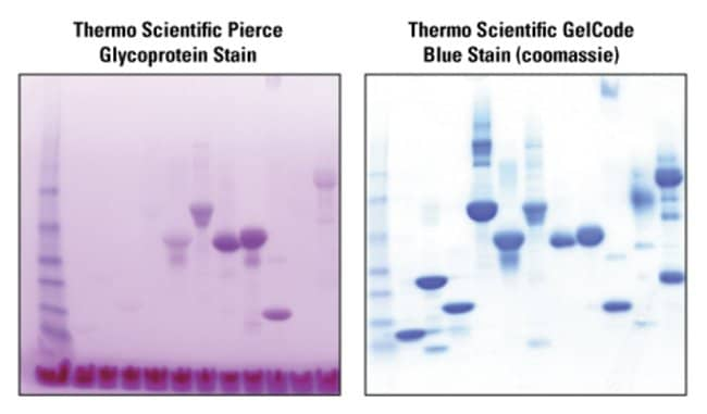
Pierce™ Glycoprotein Staining Kit
Gel Electrophoresis: Basics & Steps - SchoolWorkHelper Basic Steps. Aragonese and the buffer are mixed together and microwaved to create the gel. It is poured into a mold and has a "comb" placed in it to make holes for the DNA to be inserted. Once it has cooled the comb is removed. The gel is then placed in the gel electrophoresis box and buffer solution is poured onto it.

How to understand Gel Electrophoresis results 1 - YouTube
3 Ways to Read Gel Electrophoresis Bands - wikiHow Hold a UV light up to the gel sheet to reveal results when using a UV-based dye. With your gel sheet in front of you, find the switch on a tube of UV light to turn it on. Hold the UV light 8-16 inches (20-41 cm) away from the gel sheet. Illuminate the DNA samples with the UV light to activate the dye and read the results.
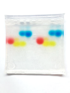
DNA101 Lab: Rainbow Gel Electrophoresis – miniPCR
How can I modify a photograph of gel electrophoresis taken with ... After amplifying my target gene (1900bp) into the required cDNA by PCR,>cut specific gel bands and purify the gel and the concentration was 30ng/ul. Next, performed A-Tailing according to the Kit...

Forensics - Science Olympiad Student Center Wiki
Part 2: Analyzing and Interpreting (Agarose) Gel Electrophoresis Results Now let see some real images. The gel image above is the result of restriction digestion. Lane 3, 5, 7, and 8 are a homozygous normal allele with a 184bp band here one band of 68bp is also present, but it is not visible. Lane 2 is a mutant uncut allele of 252bp. Lane 1 and 6 are heterozygous contain three alleles: 252bp, 184bp and 68bp.
electrophoresis.html 20_08GelElectrophoresis.jpg
Activity 2 - Gel Electrophoresis of Dyes - APS Home Remove the tape from the ends of the gel tray. Place gel into electrophoresis unit. Add 150 ml 1X TBE buffer to completely fill the box and to cover the top gel surface with about 2 mm of buffer. Note: At this point the gel box can be covered and left until the next day if necessary.
Post a Comment for "40 how to label gel electrophoresis images"