40 microscope with label
Microscope Labeling Game - PurposeGames.com About this Quiz. This is an online quiz called Microscope Labeling Game. There is a printable worksheet available for download here so you can take the quiz with pen and paper. This quiz has tags. Click on the tags below to find other quizzes on the same subject. Science. 22 Parts Of a Microscope With Their Function And Labeled Diagram The field diaphragm control is located around the lens located in the base. Hinge Screw -This screw fixes the arm to the base and allow for the tilting of the arm. Stage Clips - They hold the slide firmly onto the stage. On/OFF Switch - This switch on the base of the microscope turns the illuminator off and on.
Microscope Labeling - The Biology Corner Microscope Labeling Microscope Labeling 15. When focusing a specimen, you should always start with the _____________ objective. 16. When using the high power objective, only the _______________ knob should be used. 17. The type of microscope used in most science classes is the ______________ microscope. 18.
Microscope with label
Microscope Parts and Functions Microscope Parts and Functions With Labeled Diagram and Functions How does a Compound Microscope Work?. Before exploring microscope parts and functions, you should probably understand that the compound light microscope is more complicated than just a microscope with more than one lens.. First, the purpose of a microscope is to magnify a small object or to magnify the fine details of a larger ... Simple Microscope - Diagram (Parts labelled), Principle, Formula and Uses Parts of a Simple Microscope A simple microscope consists of Optical parts Mechanical parts Labeled Diagram of simple microscope parts Optical parts The optical parts of a simple microscope include Lens Mirror Eyepiece Lens A simple microscope uses biconvex lens to magnify the image of a specimen under focus. Microscope, Microscope Parts, Labeled Diagram, and Functions A microscope is a laboratory instrument used to examine objects that are too small to be seen by the naked eye. It is derived from Ancient Greek words and composed of mikrós, "small" and skopeîn,"to look" or "see".
Microscope with label. Histology and Microscope Slide Labels Microscope Slide Labels. These specialty Microscope Slide Labels and matching End Labels are available in standard (thin) or pathology (tissue high) thickness, and square or round corner (RC). Permanent adhesive holds labels in place during use and long-term storage. Sheet Form Size is 5¼" x 8". Prices are per thousand labels. Slide Label PDF Parts of a Microscope Printables - Homeschool Creations Label the parts of the microscope. You can use the word bank below to fill in the blanks or cut and paste the words at the bottom. Microscope Created by Jolanthe @ HomeschoolCreations.net. Parts of a eyepiece arm stageclips nosepiece focusing knobs illuminator stage objective lenses Labeling the Parts of the Microscope Labeling the Parts of the Microscope This activity has been designed for use in homes and schools. Each microscope layout (both blank and the version with answers) are available as PDF downloads. You can view a more in-depth review of each part of the microscope here. Download the Label the Parts of the Microscope PDF printable version here. Parts of a Microscope Labeling Activity - Storyboard That Create a poster that labels the parts of a microscope and includes descriptions of what each part does. Click "Start Assignment". Use a landscape poster layout (large or small). Search for a diagram of a microscope. Using arrows and textables label each part of the microscope and describe its function.
Sperm Under Microscope with Labeled Diagram » AnatomyLearner >> The ... So, under the microscope, you will identify the Type - A spermatogonium as follow - The spermatogonium is located at the base of the seminiferous tubules, Larger cells with an oval nucleus (cells are larger compared to other cells of the seminiferous tubule), Nucleus takes deep stain (posses deeply stained nucleoli), Microscope Types (with labeled diagrams) and Functions Simple microscope labeled diagram Simple microscope functions It is used in industrial applications like: Watchmakers to assemble watches Cloth industry to count the number of threads or fibers in a cloth Jewelers to examine the finer parts of jewelry Miniature artists to examine and build their work Also used to inspect finer details on products Microscope Labeling - The Biology Corner The labeling worksheet could be used as a quiz or as part of direct instruction where students label the microscope as you go over what each part is used for. The google slides shown below have the same microscope image with the labels for students to copy. Compound Microscope: Definition, Diagram, Parts, Uses, Working ... - BYJUS A compound microscope is defined as A microscope with a high resolution and uses two sets of lenses providing a 2-dimensional image of the sample. The term compound refers to the usage of more than one lens in the microscope. Also, the compound microscope is one of the types of optical microscopes.
Microscope Parts, Function, & Labeled Diagram - slidingmotion Microscope parts labeled diagram gives us all the information about its parts and their position in the microscope. Microscope Parts Labeled Diagram The principle of the Microscope gives you an exact reason to use it. It works on the 3 principles. Magnification Resolving Power Numerical Aperture. Parts of Microscope Head Base Arm Eyepiece Lens Parts of the Microscope with Labeling (also Free Printouts) Parts of the Microscope with Labeling (also Free Printouts) A microscope is one of the invaluable tools in the laboratory setting. It is used to observe things that cannot be seen by the naked eye. Table of Contents 1. Eyepiece 2. Body tube/Head 3. Turret/Nose piece 4. Objective lenses 5. Knobs (fine and coarse) 6. Stage and stage clips 7. Aperture Neuron under Microscope with Labeled Diagram - AnatomyLearner Neuron under Microscope with Labeled Diagram 31/03/2022 31/03/2022 by anatomylearner The structural and functional unit of the nervous system is the neuron that may easily observe under a light microscope. Neurons may vary considerably in size, shape, and other features. Observing Yeast Under The Microscope » Microscope Club Mix in a tablespoon of sugar until it dissolves completely. Or, mix one packet of active yeast with one tablespoon of sugar and one cup of warm water. Make sure that there are no lumps of yeast or sugar. Stir it and then let sit for almost an hour. It's also possible to observe the bubbling process of bread yeast.
Barcodes in the Lab | Learning Center | Dasco
Simple Microscope - Parts, Functions, Diagram and Labelling Simple Microscope - Parts, Functions, Diagram and Labelling A microscope is one of the commonly used equipment in a laboratory setting. A microscope is an optical instrument used to magnify an image of a tiny object; objects that are not visible to the human eyes. Table of Contents The common types of microscopes are: What is a Simple microscope?

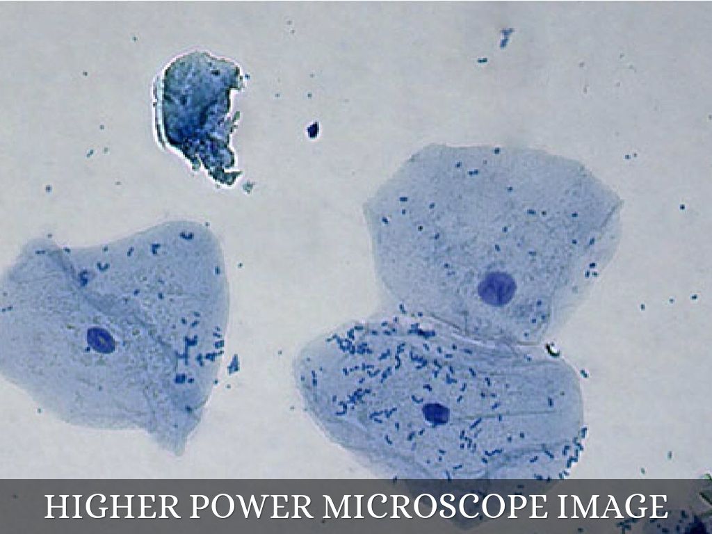
My Drawings from Lab 5 (Cells)
Quia - Label the Microscope Quiz Label the Microscope Quiz. Choose the word that correctly labels the parts of the microscope. Please enter your name. First name: Last name . Tools. Copy this to my account; E-mail to a friend; Find other activities; Start over; Print; Help; Mrs. Coyle. Simmons Elementary School. Versailles, KY: View profile;

Pennate Diatoms Under the Microscope - YouTube
Label the microscope — Science Learning Hub All microscopes share features in common. In this interactive, you can label the different parts of a microscope. Use this with the Microscope parts activity to help students identify and label the main parts of a microscope and then describe their functions. Drag and drop the text labels onto the microscope diagram.
Educational Training For Kit Epidermis Of Onion Tissue Slides Set On ...
PDF Label parts of the Microscope: Answers Label parts of the Microscope: Answers Coarse Focus Fine Focus Eyepiece Arm Rack Stop Stage Clip . Created Date: 20150715115425Z ...
TC-5400L Binocular Inverted Brightfield/Phase Contrast Biological ...
Label Microscope Diagram - EnchantedLearning.com Using the terms listed below, label the microscope diagram. arm - this attaches the eyepiece and body tube to the base. base - this supports the microscope. body tube - the tube that supports the eyepiece. coarse focus adjustment - a knob that makes large adjustments to the focus. diaphragm - an adjustable opening under the stage, allowing ...
Gastrointestinal Tract - Gallbladder Histology - Embryology
Compound Microscope Parts - Labeled Diagram and their Functions - Rs ... The eyepiece (or ocular lens) is the lens part at the top of a microscope that the viewer looks through. The standard eyepiece has a magnification of 10x. You may exchange with an optional eyepiece ranging from 5x - 30x. [In this figure] The structure inside an eyepiece. The current design of the eyepiece is no longer a single convex lens.
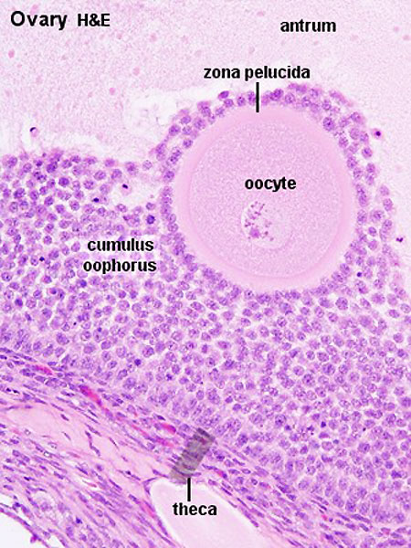
ANAT2341 Lab 1 - Oogenesis - Embryology
Explanation and Labelled Images - New York Microscope Company Another use of fluorescence imaging is Fluorescence Speckle Microscopy. It is a technology that uses fluorescence labeled macromolecular assemblies such as cytoskeletal protein to study movement and turnover rates. Fluorescence microscopy staining also is helpful in the field of mineralogical applications. It is routinely used for the study of ...

3_GeneralBio_WebCT_EntryP
Compound Microscope Parts, Functions, and Labeled Diagram Compound Microscope Definitions for Labels. Eyepiece (ocular lens) with or without Pointer: The part that is looked through at the top of the compound microscope. Eyepieces typically have a magnification between 5x & 30x. Monocular or Binocular Head: Structural support that holds & connects the eyepieces to the objective lenses.
Cells Under A Microscope by Jaimarie Nelson
Parts of a microscope with functions and labeled diagram Microscopic illuminator - This is the microscopes light source, located at the base. It is used instead of a mirror. It captures light from an external source of a low voltage of about 100v. Condenser - These are lenses that are used to collect and focus light from the illuminator into the specimen.
Post a Comment for "40 microscope with label"