41 label the features of the stomach and nearby regions in this frontal section of a cadaver
Lateral view of the brain: Anatomy and functions | Kenhub The frontal lobe takes up the majority of the superolateral surface, forming the most anterior portion of the cerebrum. It lies just deep to the frontal bone. The frontal lobe is separated from the parietal lobe by the central sulcus (fissure of Rolando), while the frontal and parietal lobes are separated from the temporal lobe by the lateral (Sylvian) sulcus. Cadaver with pictures Flashcards - Quizlet Start studying Cadaver with pictures. Learn vocabulary, terms, and more with flashcards, games, and other study tools. ... Pyloric region of stomach - narrow end of stomach. Small intestine. The last section of the digestive system, where water is absorbed from food and the remaining material is eliminated from the body, Digestive organ where ...
PDF The Cardiovascular System - Pearson nal anterior view and a frontal section. As the ana-tomical areas of the heart are described in the next section, keep referring to Figure 11.3 to locate each of the heart structures or regions.) Chambers and Associated Great Vessels Learning Objectives Trace the pathway of blood through the heart. Compare the pulmonary and systemic circuits.

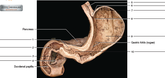
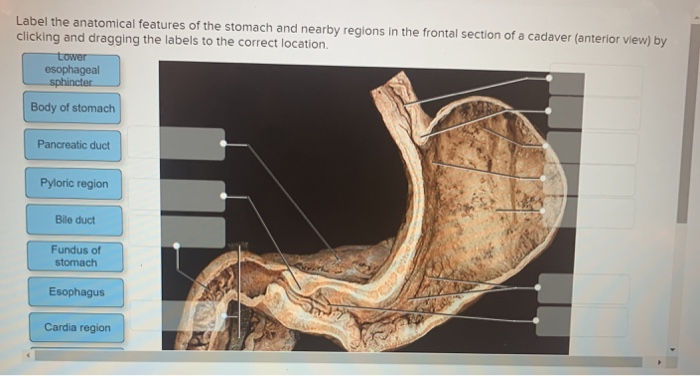
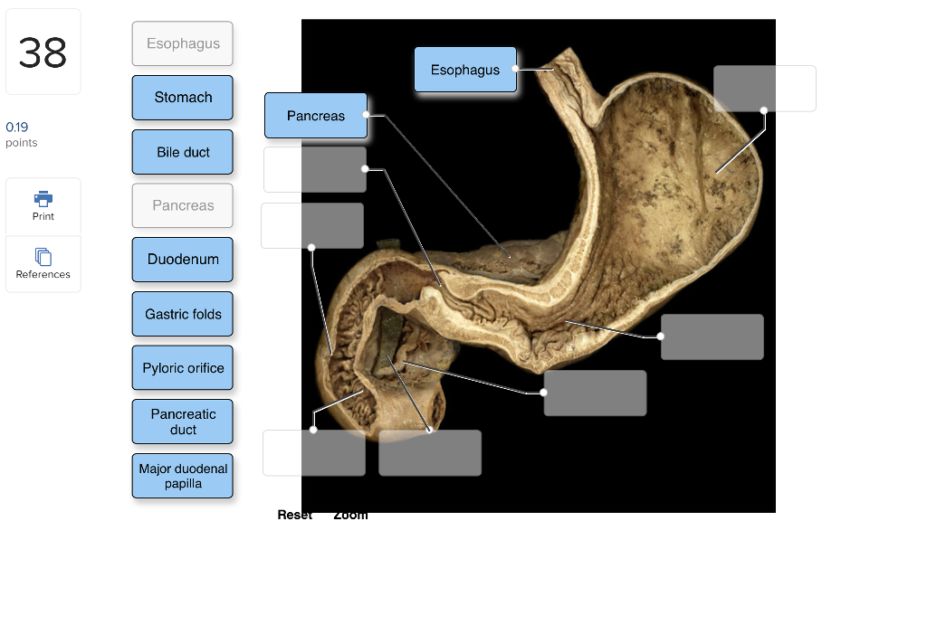
Label the features of the stomach and nearby regions in this frontal section of a cadaver
Abdomen Anatomy, Area & Diagram | Body Maps - Healthline The major muscles of the abdomen include the rectus abdominis in front, the external obliques at the sides, and the latissimus dorsi muscles in the back. The major organs of the abdomen include the... Body Cavities and Membranes: Labeled Diagram, Definitions Ventral (Anterior) and Dorsal (Posterior) Definitions: Ventral means front or toward the front of the body, and dorsal means back or toward the back of the body. We also learned in the medical prefix lecture that " ventri- " means stomach, abdomen, toward the front, or the anterior aspect of the body. Serosa four layers of the wall fig 5416 the mucus - Course Hero Region of stomach near lower esophageal sphincter 5. Contains blood vessels and nerves in a tooth 6. Responsible for peristaltic waves 7. Allow stomach to expand 8. Increase surface area for digested food absorption 9. Attached to gallbladder 10.Portion of tooth projecting beyond gingivae 11. 1. Papillary muscles 2. Tricuspid valve 3.
Label the features of the stomach and nearby regions in this frontal section of a cadaver. Vocabulary - Human Anatomy Lab Manual Primary sensory cortex - postcentral gyrus. Primary motor cortex - precentral gyrus. Primary visual cortex - occipital lobe. Primary auditory cortex - temporal lobe. Broca's speech area - for making speech. Wernicke's area - for understanding speech. Cranial Nerves and their function. I. Olfactory - sensory. II. Label the features of the stomach and nearby regions in this frontal ... The stomach is divided into four parts: 1-The cardia (8) - this part is connected to the esophagus and its where the epithelium changes from stratified squamous to columnar. In this region is the lower esophageal sphincter (6). 2--The fundus (7)- It's formed by the upper curvature of the stomach. 3- the body (9)- is the main part; and the biggest Abdominal wall: Layers, muscles and fascia | Kenhub The rectus abdominis muscles are a pair of long, straight muscles which run vertically on either side of the anterior abdominal wall. They are separated by the linea alba. The term rectus abdominis means "straight abdominal" in Latin, indicating that the muscle fibers run in a straight vertical line through the abdominal region of the body. Human Anatomy & Physiology Laboratory Manual Main Version 10th Edition ... PAL 3.0 features: • An interactive cadaver that allows students to peel back layers of the human cadaver and view hundreds of brand-new dissection photographs specifically commissioned for this version. ... A sagittal or frontal plane section of any nonspherical object, be it a banana or a body organ, provides quite a different view than a ...
The Nasal Cavity - Structure - Vasculature - TeachMeAnatomy The frontal, maxillary and anterior ethmoidal sinuses open into the middle meatus. The location of this opening is marked by the semilunar hiatus, a crescent-shaped groove on the lateral walls of the nasal cavity. The middle ethmoidal sinuses empty out onto a structure called the ethmoidal bulla. This is a bulge in the lateral wall formed by ... Solved 11 Label the anatomical features of the stomach and - Chegg question: 11 label the anatomical features of the stomach and nearby regions in the frontal section of a cadaver (anterior view) by clicking and dragging the labels to the correct location gastric folds (ruga) 1 points duodenum pancreas references pancrew duct esophagus body of stomach pylone region pylon sphinchi cardia region lewer esophageal … Digestive System Anatomy, Area, and Diagram | Body Maps The largest parts of the digestive system include: Esophagus: A hollow tubular organ in the neck and chest area that connects the mouth to the stomach. Muscles here propel food to the stomach ... Female Body Diagram: Parts of a Vagina, Location, Function Clitoris: The clitoris sits at the top of the vulva, above the urethral opening. A fold of skin called the clitoral hood covers most of the clitoris, leaving only the tip or nub visible. The rest of the clitoris is a spongy shaft that goes back several inches inside the body.
Illustrated Anatomy of the Stomach - ThoughtCo The stomach is an organ of the digestive system. It is an expanded section of the digestive tube between the esophagus and small intestine. Its characteristic shape is well known. The right side of the stomach is called the greater curvature and the left the lesser curvature. PDF Body Organization and Terminology ASSIGNMENT Human Cadaver section, click on the appropriate organ system, then click on Show Labels and find: a. External abdominal oblique muscle (Muscular System) b. Sciatic nerve (Nervous System) c. Pancreas and Thyroid gland (Endocrine System) d. Inferior Vena Cava (Cardiovascular System) e. Lymph Vessel (Lymphatic System) f. Axial Skeleton (80 bones) | SEER Training Vertebral Column. Cervical vertebrae (7) Thoracic vertebrae (12) Lumbar vertebrae (5) Sacrum (1) Coccyx (1) Lumen inferior end 544 figure 5411 normalappendix 2 - Course Hero Locate the four layers of the wall (fig. 54.13). The mucus functions as a lubrication and holds the particles of fecal matter together. 5. Prepare a labeled sketch of the wall of the large intestine in Part A of the laboratory assessment. 6. Complete Parts B, C, and D of the laboratory assessment.
Solved: PART B: Assessments Identify The Numbered Features... | Chegg.com
Lab 8: Digestive System - Human Anatomy Lab Manual The peritoneum is a large serous membrane which lines the abdominal cavity and coverers most of the digestive organs. some organs are only partially covered by the peritoneum while others are entirely uncovered. These organs are referred to as being retroperitoneal.
Solved: Identify the numbered features in figures 54.14, 54.15 ...
The Abdomen (Human Anatomy) - Picture, Function, Parts ... - WebMD In the front, the abdomen is protected by a thin, tough layer of tissue called fascia. In front of the fascia are the abdominal muscles and skin. In the rear of the abdomen are the back muscles and...
Solved: Label The Anatomical Features Of The Stomach And N... | Chegg.com
Anatomical Position: Body Planes and Sections - EZmed Sagittal Section: A sagittal section is created when a cut is made down the sagittal plane, providing a side view of the body. Coronal (Frontal) Plane Next we have the coronal plane. Remember the coronal plane was the "C" in our abbreviation "SCT", which helps us remember the 3 major planes of the body.
Solved: Label The Internal Features Of Stomach And Duodenu... | Chegg.com
The Trachea (Human Anatomy): Picture, Function, Conditions, and ... - WebMD The trachea, commonly known as the windpipe, is a tube about 4 inches long and less than an inch in diameter in most people. The trachea begins just under the larynx (voice box) and runs down ...
Post a Comment for "41 label the features of the stomach and nearby regions in this frontal section of a cadaver"