45 anterior view of the heart with labels
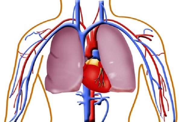
Understanding an ECG | ECG Interpretation | Geeky Medics An ECG lead is a graphical representation of the heart's electrical activity which is calculated by analysing data from several ECG electrodes. Chest leads. V1: septal view of the heart; V2: septal view of the heart; V3: anterior view of the heart; V4: anterior view of the heart; V5: lateral view of the heart; V6: lateral view of the heart ... Anatomy Tutorial - Anterior | Atlas of Human Cardiac Anatomy This illustration demonstrates an anterior view of the thoracic cavity, highlighting the position of the heart in relationship to the ribs and diaphragm. The right atrium, right ventricle, and a small portion of the left ventricle are visible from this aspect.
Heart Anatomy - Anterior (Front) View : Medical Illustration This medical exhibit pictures an anterior (front) view of the heart anatomy with labels for the aorta, superior vena cava, right atrium, right ventricle, inferior vena cava, pulmonary trunk, left atrium, pulmonary veins and left ventricle. Max Image Size: 2448 pixels wide by 1804 pixels high. Recent Comments. No comments have been posted.
Anterior view of the heart with labels
Lab Report 38 Figures 38.1, 38.2, and 38.3.pdf - Course Hero Figure 38.1- Label this anterior view of the human heart. 1.) Aorta 2.) Left pulmonary artery 3.) Left pulmonary veins 4.) Left atrium 5.) Left ventricle 6.) Apex 7.) Superior vena cava 8.) Right atrium 9.) Inferior vena cava 10.) Right ventricle Aorta Left pulmonary artery Left pulmonary veins Left atrium Left ventricle Apex Superior vena cava Right atrium Inferior vena cava Right ventricle anterior heart Quiz - PurposeGames.com This is an online quiz called anterior heart There is a printable worksheet available for download here so you can take the quiz with pen and paper. From the quiz author quiz game to assist a&p students with anatomy of exterior heart. This quiz has tags. Click on the tags below to find other quizzes on the same subject. quiz heart label anterior posterior view of heart labeled - polarisheartandvascular A Anterior view of the external heart C 2019 Pearson Education. Label the 4 chambers as well as the major vessels entering and leaving these chambers. Your heart is the main organ of your cardiovascular system a network of blood vessels that pumps blood throughout your body. This quiz has tags. Terms in this set 15 Aorta.
Anterior view of the heart with labels. Anatomy of the heart and coronary arteries (coronary CT) - IMAIOS Anatomy of the human heart and coronaries: how to view anatomical structures. This tool provides access to an MDCT atlas in the 4 usual planes, allowing the user to interactively discover the heart anatomy. The images are labeled, providing an important medical and anatomical tool. The quiz mode makes it possible to evaluate the user's progress. Solved Label the structures seen in the anterior view of the - Chegg View the full answer Transcribed image text: Label the structures seen in the anterior view of the heart. Superior vena cava Interior vena cava Aorta Left atrium Pulmonary trunk Pulmonary vein Left ventricle Pulmonary artery Right ventricle Right atrium Previous question Next question 19.1 Heart Anatomy - Anatomy & Physiology The position of the heart in the torso between the vertebrae and sternum (see Figure 19.1.1 for the position of the heart within the thorax) allows for individuals to apply an emergency technique known as cardiopulmonary resuscitation (CPR) if the heart of a patient should stop. By applying pressure with the flat portion of one hand on the sternum in the area between the line at T4 and T9 ... Label the Heart Diagram Anterior view Quiz - PurposeGames.com Jan 26, 2017 · This is an online quiz called Label the Heart Diagram Anterior view There is a printable worksheet available for download here so you can take the quiz with pen and paper. Your Skills & Rank Total Points 0 Get started! Today's Rank -- 0 Today 's Points One of us! Game Points 24 You need to get 100% to score the 24 points available Actions 2 favs
Heart - Collection Page | AnatomyTOOL 3D model of the normal heart and lungs with numbered English labels. From a collaboration of Universities of Leiden, Delft and Groningen. ... A photo of the anterior view of a plastinated normal heart, where highlights on structures can be switched on and off, by Univ. of Br. Columbia. Anterior View Human Body Illustrations & Vectors Rib Cage of Human Skeleton System Anatomy with detailed labels Anterior View. 3D Illustration Concept of Rib Cage of Human Skeleton System Anatomy with detailed. Human body front, back and side views. ... The human heart anatomy Anterior View, Frontal section and cardiac muscle structure. Spine Vertebrae - Lateral view / Side view. Human body ... Human Heart-Anterior View - John Burroughs School Frontal Heart Anatomy. Untitled Document. Label the anterior view of the human heart. The correct name of the structure will appear below when you click on the number of the structure. A hand appears when the pointer is positioned properly over the number. Click the mouse when the hand appears. Go ahead, start with number 1. Heart Labeling anterior view Diagram - Quizlet Heart Labeling anterior view STUDY Learn Write Test PLAY Match + − Created by Meghan12th PLUS Terms in this set (26) brachiocephalic trunk ... left common carotid artery ... superior vena cava ... aortic arch ... liigamentum arteriosum ... right pulmonary artery ... amending aorta ... right pulmonary veins ... pulmonary trunk ... right atrium ...
Heart Anatomy: Labeled Diagram, Structures, Blood Flow ... - EZmed There are 3 tricks to remember the tricuspid valve is located on the right side of the heart, and the mitral valve is located on the left. Trick 1: You can use the saying "TRI (Try) it before you BI (buy) it". This will help you remember the tricuspid valve comes before the bicuspid/mitral valve. Anterior External View of Heart Labeling Diagram | Quizlet Anterior External View of Heart Labeling Diagram | Quizlet Anterior External View of Heart Labeling STUDY Learn Write Test PLAY Match Created by randajsmith Terms in this set (14) Superior Vena Cava ... Superior Vena Cava (SVC) ... Right Pulmonary Artery ... Right Pulmonary Veins ... Right Atrium ... Right Coronary Artery ... Right Ventricle ... Heart Anatomy: Heart Dissection - University of Washington The picture below shows an anterior view of the heart with the pericardium removed. The letters indicated in the text refer to the labels on the picture. The anterior surface of the heart is characterized by the presence of the large arteries leaving the base of the heart, the pulmonary trunk (H) and the aorta (G). Heart Anterior View Stock Photos and Images - Alamy Anterior (Front) View of the Heart ID: ADTXR9 (RM) Human Body Organs Label Design Anterior view Anatomy ID: KYXGRX (RF) Vintage anatomy print features a dual view of the human heart. ID: 2A13W0N (RF) Front view of the heart and it's coronary arteries ID: GD1N7W (RF) Anterior view of heart and lung on green background. ID: E6BJB3 (RM)
Heart Model Contiunued - ProProfs Quiz
Heart chambers and associated great vessels - Anatomy Image: Anterior view of the human heart with labels. right atrium has two basic parts: a smooth posterior and an anterior portion in which bundles of muscle tissue form ridges in the walls. The muscle bundles are called pectinate muscles because they look like the teeth of a comb.

Pedi cardiology: Pericardial reflections
Solved Heart Chambers and Valves: Frontal Heart Section 2 - Chegg Heart Chambers and Valves: Frontal Heart Section 2 Label the chambers and valves seen in an anterior view of the heart Left ventricle Right ventricle Aortic valve Tricuspid Valve Right atrum Mihral valve Chord Pulmonary ave
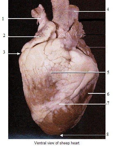
BIO202-Sheep Heart
Blausen 0451 - Anterior view of the heart - English labels Anterior view of the heart. English labels. By Blausen Medical Communications, Inc. Retrieved from Wikimedia Commons: category: Images from Blausen Medical Communications. Fig. 0451. Anatomical structures in item: Cor. Auricula dextra. Auricula sinistra. Truncus pulmonalis. Arcus aortae.

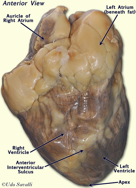
Lab 2 Pig Heart-Labeled | Cardiovascular, Estudos
Heart Anatomy - Interior View - Maricopa Other Cardiac Tutorials... Internal Heart Tutorial and Self-Test to review basic anatomy of heart, chambers, valves etc... Posterior Heart Tutorial and Self-Test to review basic anatomy of heart and vessels... Quizzer 1 on basic EKG features including deflections and segments etc... EKG Arrhythmia Tutorial to identify arrhythmias including VTAC, PVC, hyperkalemia etc...

Heart Anatomy | Anatomy and Physiology II
Heart Diagram with Labels and Detailed Explanation - BYJUS Diagram of Heart. The human heart is the most crucial organ of the human body. It pumps blood from the heart to different parts of the body and back to the heart. The most common heart attack symptoms or warning signs are chest pain, breathlessness, nausea, sweating etc. The diagram of heart is beneficial for Class 10 and 12 and is frequently ...

Brenda's A & P Eportfolio: Objective 23 & 24: Vessels and Structure of ...
Diagrams, quizzes and worksheets of the heart | Kenhub Labeled heart diagrams Take a look at our labeled heart diagrams (see below) to get an overview of all of the parts of the heart. Once you're feeling confident, you can test yourself using the unlabeled diagrams of the parts of the heart below. Labeled heart diagram showing the heart from anterior Unlabeled heart diagrams (free download!)

Anterior interventricular coronary artery - The Anatomy of the Heart ...
Circulatory System Coloring and Labeling - Lovejoy Anatomy and ... - Google SUPERIOR ASPECT OF THE HEART This view of the heart is seen as if the atria and the major vessels have been removed. You should be able to see all of the major valves of the heart. The most anterior valve is the pulmonary semilunar valve that occurs between the right ventricle and the pulmonary trunk. Label and color this valve blue.Posterior to this is the aortic semilunar valve.

Cardiac Chambers: The Four-Chamber and Short-Axis Views | Obgyn Key
Heart: illustrated anatomy - e-Anatomy - IMAIOS This interactive atlas of human heart anatomy is based on medical illustrations and cadaver photography. The user can show or hide the anatomical labels which provide a useful tool to create illustrations perfectly adapted for teaching. Anatomy of the heart: anatomical illustrations and structures, 3D model and photographs of dissection.
Free Heart Diagram Unlabeled, Download Free Heart Diagram Unlabeled png ...
Human Heart - Diagram and Anatomy of the Heart - Innerbody The heart is a muscular organ about the size of a closed fist that functions as the body's circulatory pump. It takes in deoxygenated blood through the veins and delivers it to the lungs for oxygenation before pumping it into the various arteries (which provide oxygen and nutrients to body tissues by transporting the blood throughout the body).
Post a Comment for "45 anterior view of the heart with labels"