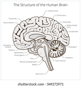
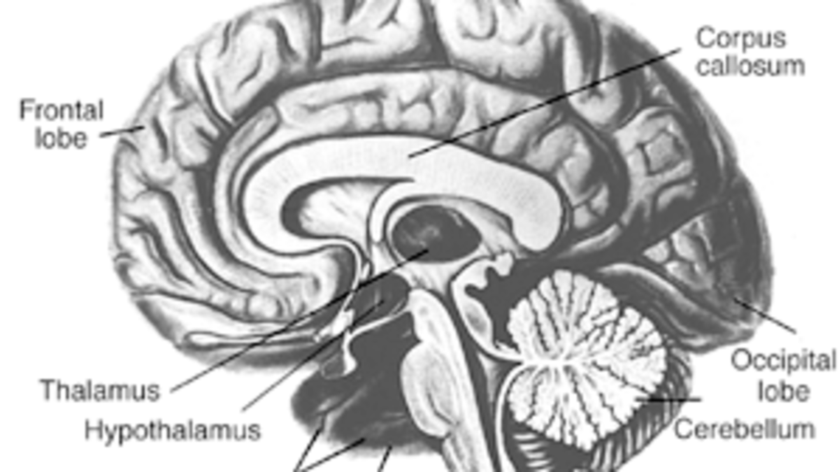
44 brain fully labeled
When the Brain Starts Adulting When the Brain Starts Adulting. Your brain changes a lot between birth and adolescence. It grows in overall size, modifies the number of cells contained within, and transforms the degree of connectivity. The changes don't stop once you turn 18. In fact, scientists now think your brain continues maturing and fine-tuning itself well into your 20s. CT head sagittal - labeling questions | Radiology Case - Radiopaedia The labeled structures are (excluding the correct side): mastoid air cells temporalis muscle zygomatic arch external auditory (acoustic) canal temporomandibular joint tentorium cerebelli sylvian fissure temporal lobe occipital lobe carotid canal trigone (atrium) of the lateral ventricle centrum semiovale forceps minor frontal lobe
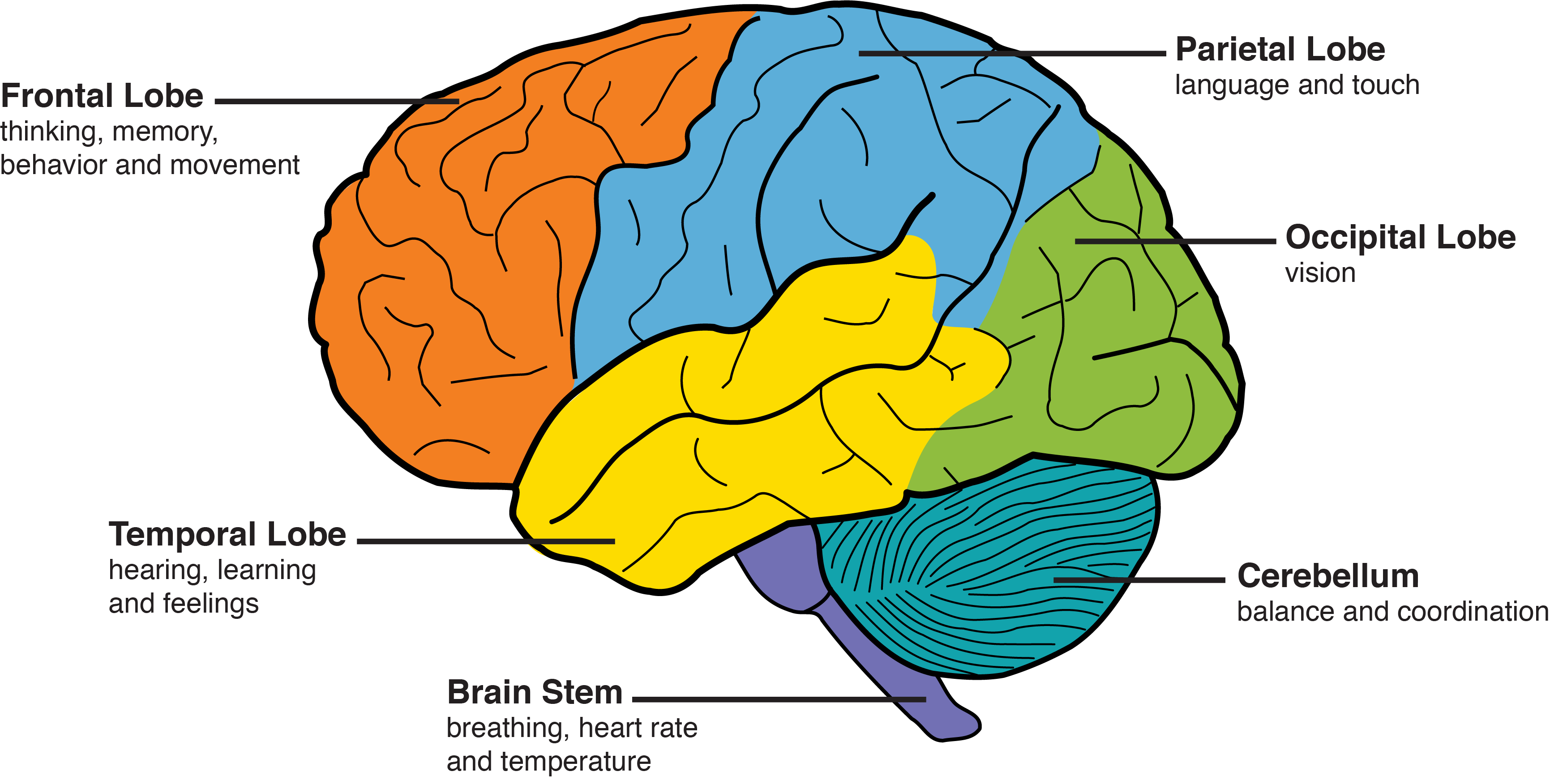
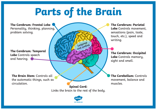
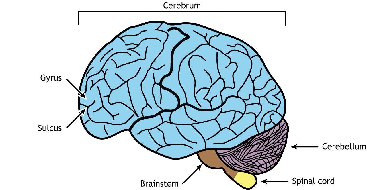
Labeled Brain Model Diagram - Science Trends The brainstem is found at the very bottom of the brain, connecting the rest of the brain to the spinal column. It is the oldest region of the brain in terms of the brain's evolution. The brainstem is responsible for the regulation of actions necessary for survival such as breathing, sleep, digestion, heart rate, and reflexes.
Brain fully labeled
Human brain - Wikipedia The human brain is the central organ of the human nervous system, and with the spinal cord makes up the central nervous system.The brain consists of the cerebrum, the brainstem and the cerebellum.It controls most of the activities of the body, processing, integrating, and coordinating the information it receives from the sense organs, and making decisions as to the instructions sent to the ... Right Brain, Left Brain: A Misnomer | Dana Foundation August 2, 2019 The human brain is basically symmetrical, split down the middle: the right cerebral hemisphere receives sensory input from and directs movement on the left side of the body, while the left hemisphere governs corresponding functions for the right side. Symmetry only goes so far, however—there are differences, too. NIMH » Publications About Brain Anatomy and Physiology Get Excited about the Brain! This science education activity book intended for children ages 8-12 years old helps kids learn facts about the brain through games and puzzles about brain science and research. This activity book can be downloaded and printed. En español The Teen Brain: 7 Things to Know
Brain fully labeled. Brain Regions and Functions | Ask A Biologist The brain is a very busy organ. It is the control center for the body. It runs your organs such as your heart and lungs. It is also busy working with other parts of your body. All of your senses - sight, smell, hearing, touch, and taste - depend on your brain. Tasting food with the sensors on your tongue is only possible if the signals from ... Brain (Human Anatomy): Picture, Function, Parts, Conditions, and More The brain is made up of many specialized areas that work together: • The cortex is the outermost layer of brain cells. Thinking and voluntary movements begin in the cortex. • The brain stem is... How Does Your Stomach Tell Your Brain That You're Full? A variety of signals from your body tell your brain when your stomach is full. Satiety, the sensation that you've had enough to eat, results from a balance of hormonal and neurological signals reaching your brain from your stomach. Other factors, such as the sensory quality of food, also contribute to satiety. Sagittal Section of Human Brain with Labeled Parts stock photo ... Description 3D Computer Graphic Image with labeled side view of human brain parts on white background. 1 credit Essentials collection for this image $4 with a 1-month subscription (10 Essentials images for $40) Continue with purchase View plans and pricing Includes our standard license. Add an extended license. Credit: Hank Grebe
It's All in Your Brain: the Structure of ADHD - Healthline Researchers also looked at the differences in white and grey matter in children with and without ADHD. White matter consists of axons, or nerve fibers. Grey matter is the outer layer of the brain ... Brain Explorer :: Allen Brain Atlas: Human Brain The Brain Explorer 2 software is a desktop application for viewing the human brain anatomy and gene expression data in 3-D. Using the Brain Explorer 2 software, you can: View a fully interactive version of the Allen Human Brain Atlas in 3-D. View gene expression data in 3-D: inflated cortical surfaces are colored by gene expression values of ... Brain Basics: Understanding Sleep | National Institute of Neurological ... Norepinephrine and orexin (also called hypocretin) keep some parts of the brain active while we are awake. Other neurotransmitters that shape sleep and wakefulness include acetylcholine, histamine, adrenaline, cortisol, and serotonin. Genes and sleep Genes may play a significant role in how much sleep we need. Central nervous system: Anatomy, structure, function | Kenhub The brainstem is the inferior-most part of the brain. It sits in the posterior cranial fossa and consists of three parts: midbrain, pons and medulla oblongata. Internally, it is divided into the basal area, tegmentum and tectum. The brainstem has three main important functions ; It contains the nuclei of the majority of the cranial nerves.
Labeled Dog Brain Diagram 2022 Question By Labeled Dog Brain Diagram ... 2946147276 best questions for Labeled dog brain diagram collected 147276 best questions the«Labeled dog brain diagram» category soyou can quickly find the answer your question popular questionsWhy are certain dog breeds labeled dangerous Read... thedogvisitor.com. Trends; close Brain - Wikipedia A brain is an organ that serves as the center of the nervous system in all vertebrate and most invertebrate animals. It is located in the head, usually close to the sensory organs for senses such as vision. It is the most complex organ in a vertebrate's body. Left brain vs. right brain: Characteristics, functions, and myths The brain's left half is primarily responsible for speech and abstract thinking. It also controls the right side of the body. The right side of the brain is responsible for image processing,... 2,782 Labeled brain anatomy Images, Stock Photos & Vectors - Shutterstock Labeled brain anatomy royalty-free images 2,782 labeled brain anatomy stock photos, vectors, and illustrations are available royalty-free. See labeled brain anatomy stock video clips Image type Orientation People Artists More Sort by Healthcare and Medical Anatomy human brain brain organ medicine human body cerebellum cerebral cortex limbic system
Behavior & Personality Changes | Memory and Aging Center
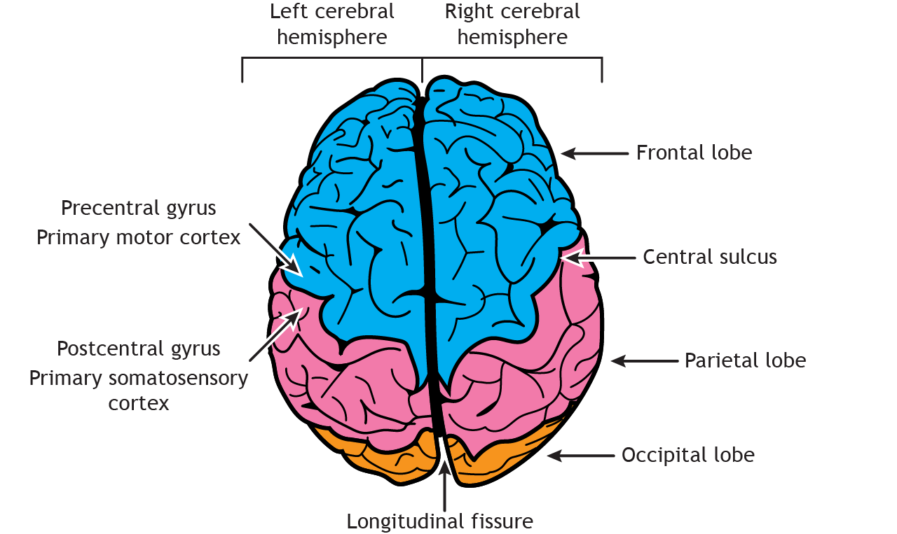
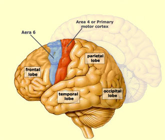
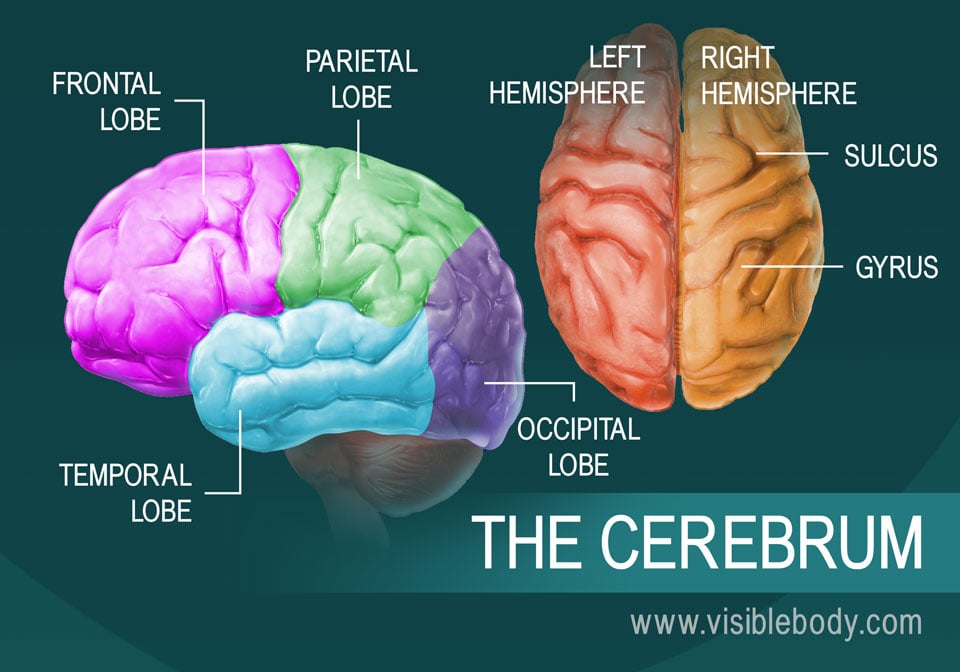
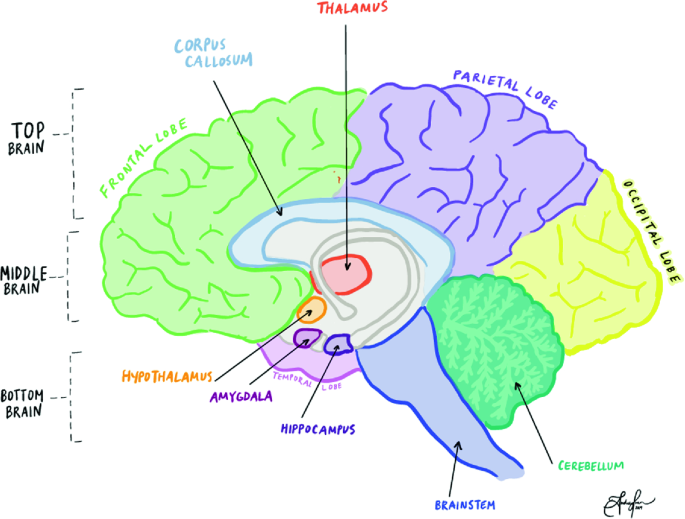
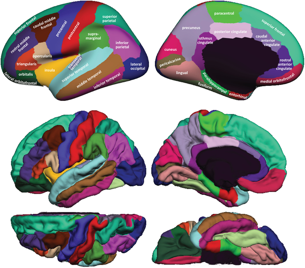
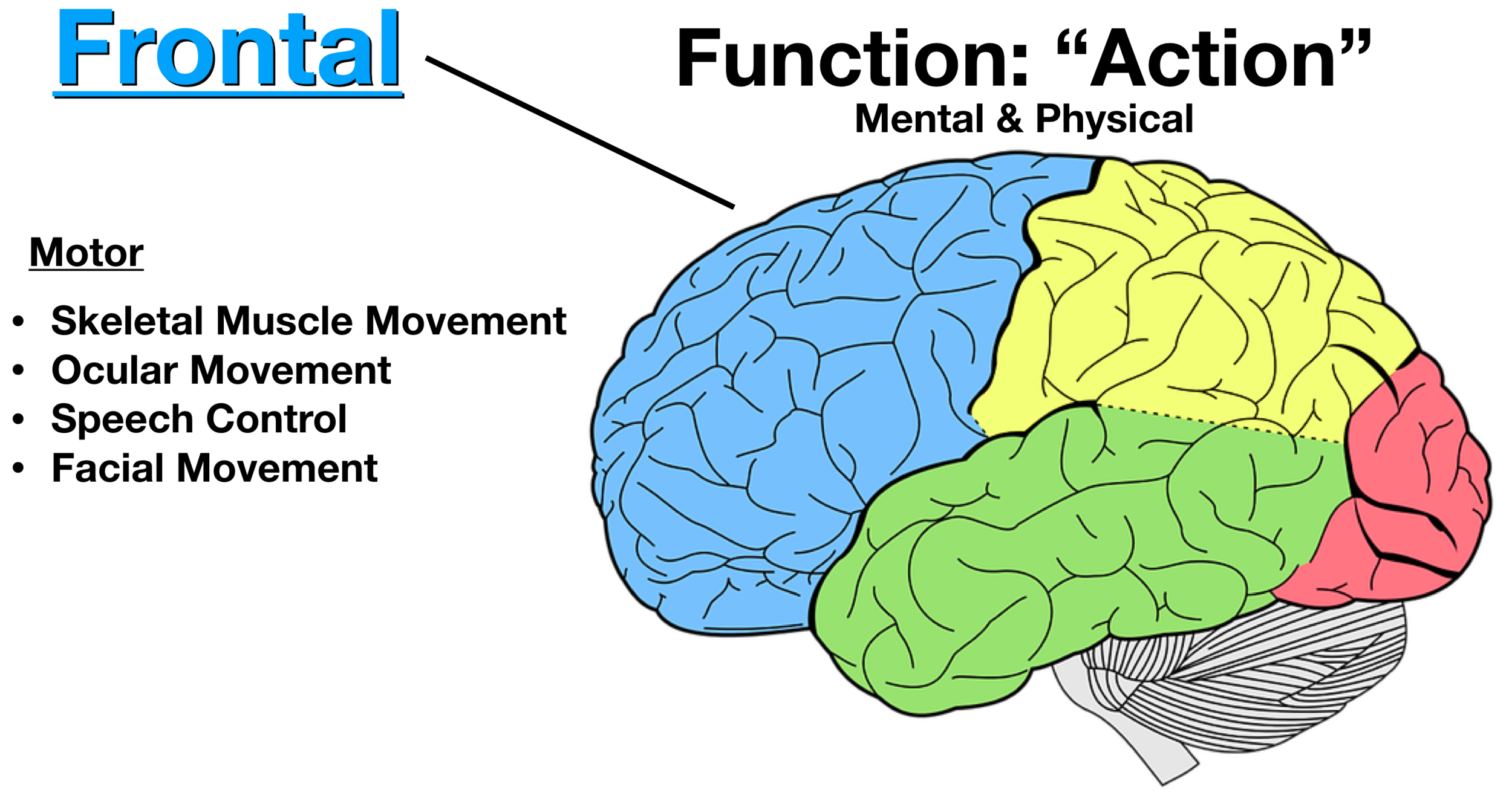
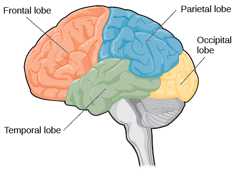
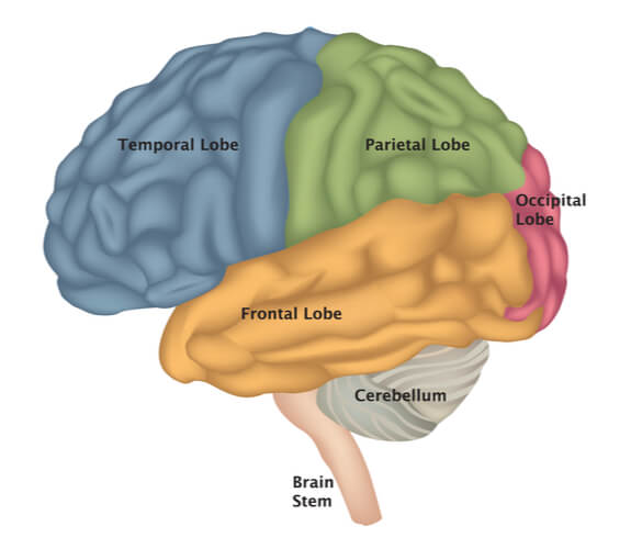
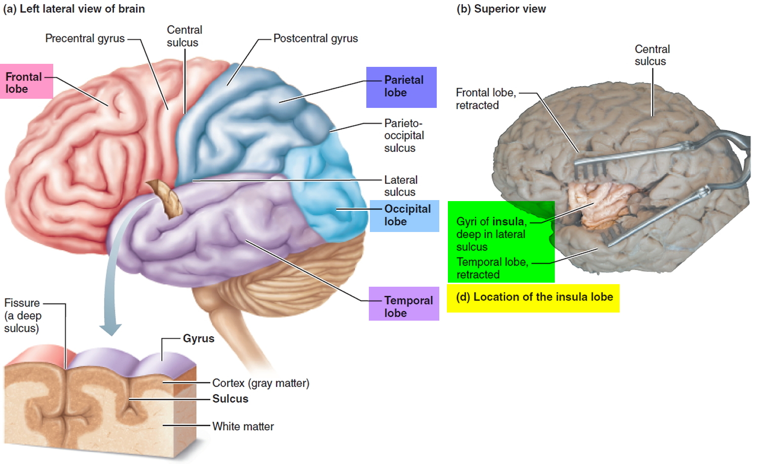
Frontal Lobe: Function, Location, and Structure - Spinal Cord The frontal lobe plays a key role in this complex set of cognitive functions. Named for its location, the frontal lobe is situated toward the front of the cerebrum, just behind the forehead and under the frontal skull bones. It sits atop the temporal lobe, in front of the parietal lobe, and apart from the occipital lobe, with portions of the ...
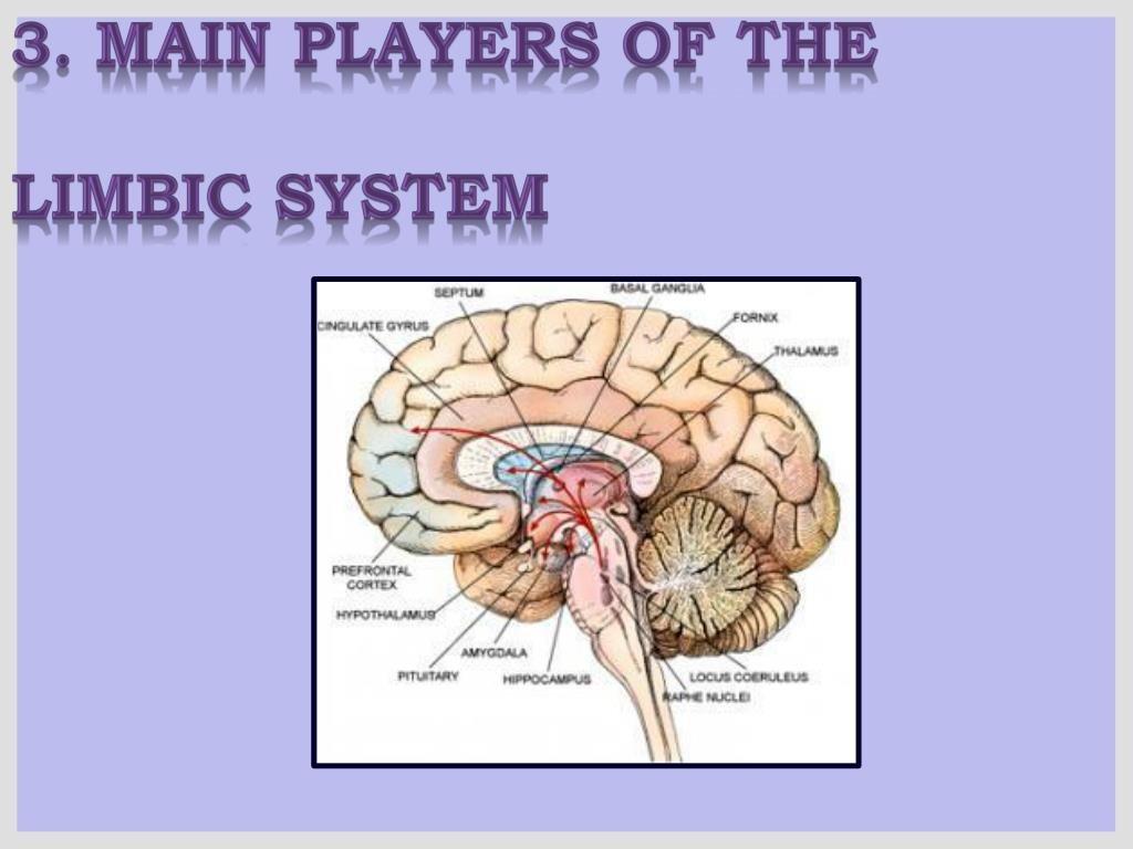
PPT - LIMBIC SYSTEM PowerPoint Presentation, free download ...
Neuroanatomy, Temporal Lobe - StatPearls - NCBI Bookshelf The temporal lobe of the brain is often referred to as the neocortex. It forms the cerebral cortex in conjunction with the occipital lobe, the parietal lobe, and the frontal lobe. It is located mainly in the middle cranial fossa, a space located close to the skull base. It is anterior to the occipital lobe and posterior to the frontal lobe. It is found inferior to the lateral fissure, also ...

How to Draw a Brain: 14 Steps (with Pictures) - wikiHow
Labeled Dog Brain Anatomy 2022 Question By Labeled Dog Brain Anatomy ... 2946147275 best questions for Labeled dog brain anatomy collected 147275 best questions the«Labeled dog brain anatomy» category soyou can quickly find the answer your question popular questionsWhy are certain dog breeds labeled dangerous Read... thedogvisitor.com. Trends; close
External Brain Anatomy – Foundations of Neuroscience
Brain Anatomy | Definition and Patient Education - Healthline The brain, along with the spinal cord is a crucial component of the central nervous system (CNS). Two types of cells exist in the brain. Neurons send and receive signals to and from your brain and ...
What is the Brain? For Kids | Information and Resources
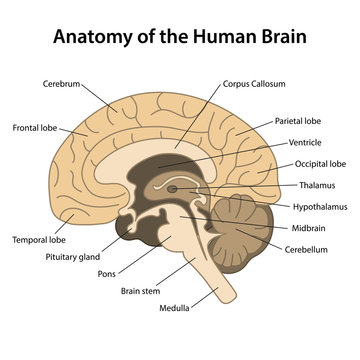
14 Informative Facts, Diagram & Parts Of Human Brain For Kids The brain and spinal cord are part of the central nervous system (CNS). The brain weighs just about two to three pounds and appears like a walnut. The brain is comprised of three main regions — cerebrum, cerebellum, and brainstem (3). Save Image: Shutterstock Let us discuss these parts and their functions in more detail (1) (3) (4).
THE BRAIN FROM TOP TO BOTTOM
Labeled Diagrams of the Human Brain You'll Want to Copy Now The average dimension of the adult human brain is 5.5 inches in width and 6.5 inches in length. The height of the human brain is about 3.6 inches and it weighs about 4 to 5 lbs at birth and 3 lbs in adults. The total surface area of the cerebral cortex is about 2,500 cm2 and when stretched, it will cover the area of a night table.
The Human Brain
5 unsolved mysteries about the brain - Allen Institute Electron microscope, or EM, images of a section of the human brain generated at the Allen Institute. This technique allows researchers to map brain tissue down to the level of its individual connections, or synapses. Biology textbooks tell us that the brain communicates via synapses, specialized connections between two different neurons.
The Developing Brain and Trauma | SpringerLink
Labeled Dog Brain 2022 Question By Labeled Dog Brain - The Dog Visitor Labeled dog brain - Page 1/2946 147275 best questions for Labeled dog brain We've collected 147275 best questions in the « Labeled dog brain » category so you can quickly find the answer to your question!

Labelled Diagram Stock Illustrations – 315 Labelled Diagram ...
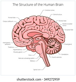
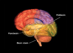
Brain Basics: Know Your Brain | National Institute of Neurological ... The brain can be divided into three basic units: the forebrain, the midbrain, and the hindbrain. The hindbrain includes the upper part of the spinal cord, the brain stem, and a wrinkled ball of tissue called the cerebellum ( 1 ). The hindbrain controls the body's vital functions such as respiration and heart rate.
Frontiers | 101 Labeled Brain Images and a Consistent Human ...
Parts of the Human Brain | Anatomy & Function - Study.com The parts of the brain include the cerebrum, the cerebellum, the brain stem, and the pituitary gland. The brain structure is protected by the skull, which is composed of the cranium and the bones...
Draw a labelled diagram of human brain and mention the ...
NIMH » Publications About Brain Anatomy and Physiology Get Excited about the Brain! This science education activity book intended for children ages 8-12 years old helps kids learn facts about the brain through games and puzzles about brain science and research. This activity book can be downloaded and printed. En español The Teen Brain: 7 Things to Know
Lobes of the Brain: Cerebral Cortex Anatomy, Function ...
Right Brain, Left Brain: A Misnomer | Dana Foundation August 2, 2019 The human brain is basically symmetrical, split down the middle: the right cerebral hemisphere receives sensory input from and directs movement on the left side of the body, while the left hemisphere governs corresponding functions for the right side. Symmetry only goes so far, however—there are differences, too.
8,770 Cerebral cortex Images, Stock Photos & Vectors ...
Human brain - Wikipedia The human brain is the central organ of the human nervous system, and with the spinal cord makes up the central nervous system.The brain consists of the cerebrum, the brainstem and the cerebellum.It controls most of the activities of the body, processing, integrating, and coordinating the information it receives from the sense organs, and making decisions as to the instructions sent to the ...

PPT – The Biological Basis for Behavior PowerPoint ...

brain parts Diagram | Quizlet

How to draw human brain/ draw labelled diagram of brain/brain diagram/draw and label brain diagram

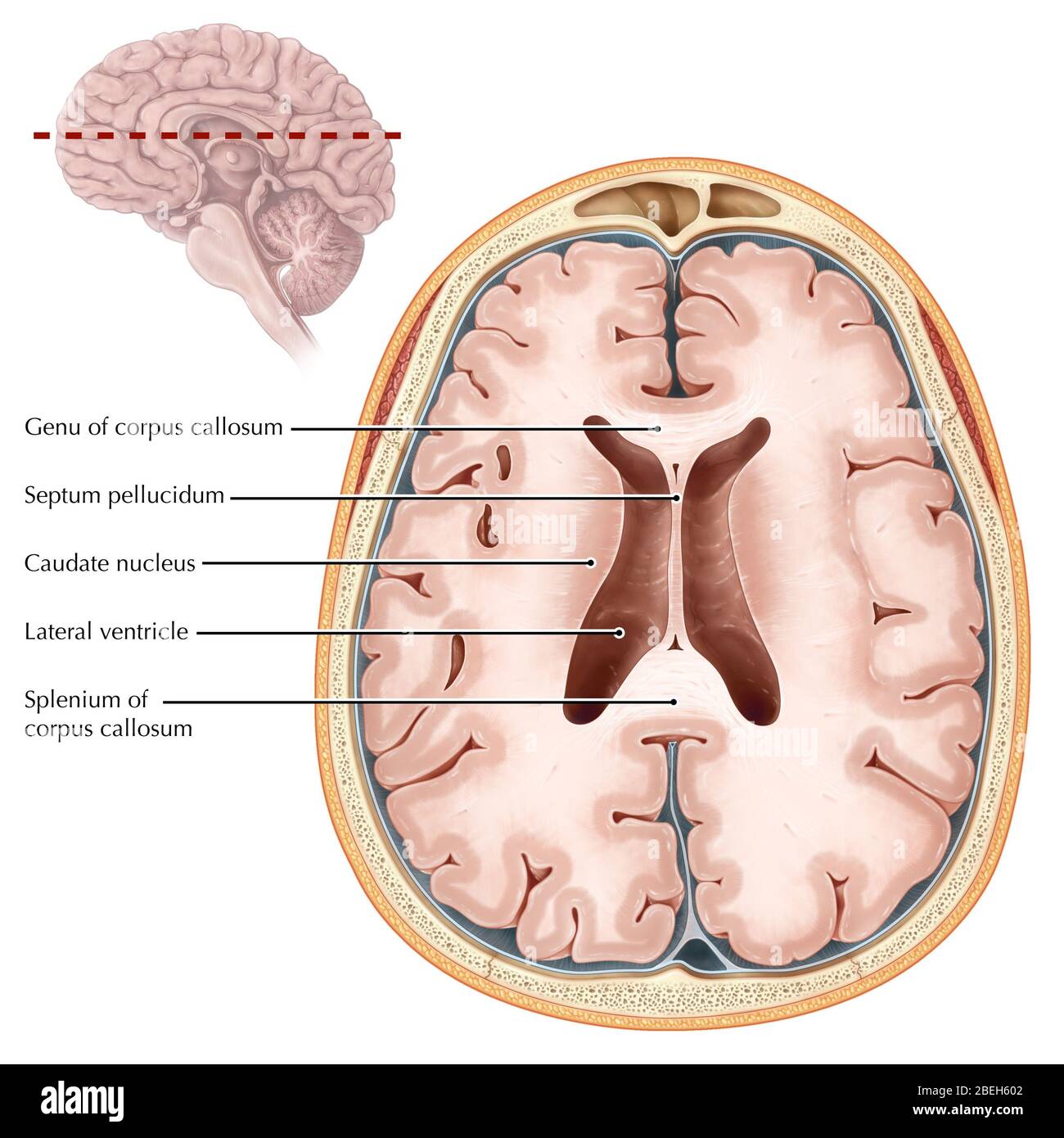
Duke Neurosciences - Lab 5: Forebrain Sectional Anatomy

Brain is not fully mature until 30s and 40s
External Brain Anatomy – Foundations of Neuroscience

Ventricles of the Brain: Labeled Anatomy, Function, CSF Flow ...

Pin on Frontotemporal Dementia

Lobes of the Brain: Cerebral Cortex Anatomy, Function ...

Cortical homunculus Korteks serebral Pemetaan otak Sistem ...
Lobes of the Brain | Introduction to Psychology

Brain Facts: The Four Lobes
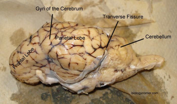
Sheep Brain Dissection with Labeled Images
2,783 Labeled brain anatomy Images, Stock Photos & Vectors ...
Labeled Brain Images – Browse 701 Stock Photos, Vectors, and ...
Brain Stem Stroke | American Stroke Association

The Amygdala by Borbála Kis
2,783 Labeled brain anatomy Images, Stock Photos & Vectors ...
Brain Stem - The Definitive Guide | Biology Dictionary
Wet Brain: Thiamine, Diet, and Abstinence - HealthProAdvice

The Brain - Diagram and Explanation
Psychology – Brain Structure/Anatomy and Function
Human Brain Anatomy and Function - Cerebrum, Brainstem

The Brain (5.1.4) | AQA GCSE Biology Revision Notes 2018 ...

File:Dopaminergic pathways.svg - Wikimedia Commons

Duke Neurosciences - Lab 5: Forebrain Sectional Anatomy

Free brain - Vector Art

Brain Facts: The Four Lobes
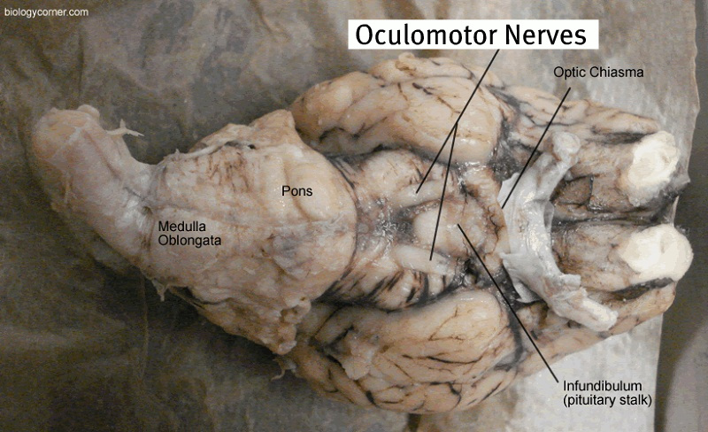
Sheep Brain Dissection with Labeled Images
:max_bytes(150000):strip_icc()/medial_view_brain-571823d23df78c3fa2be9e2d.jpg)
Divisions of the Brain: Forebrain, Midbrain, Hindbrain
Brain transverse section hi-res stock photography and images ...
Post a Comment for "44 brain fully labeled"