41 eye labeling diagram
Eye diagram labeling - Printable - PurposeGames.com Eye diagram labeling - Printable Download and print this quiz as a worksheet. You can modify it to fit your needs before you download. This is a printable worksheet made from a PurposeGames Quiz. To play the game online, visit Eye diagram labeling Download Printable Worksheet Please note! Labelling the eye — Science Learning Hub Labelling the eye — Science Learning Hub Activity Labelling the eye The human eye contains structures that allow it to perceive light, movement and colour differences. In this activity, students use online or paper resources to identity and label the main parts of the human eye. Citizen science Teacher PLD Glossary Sign in Email Us
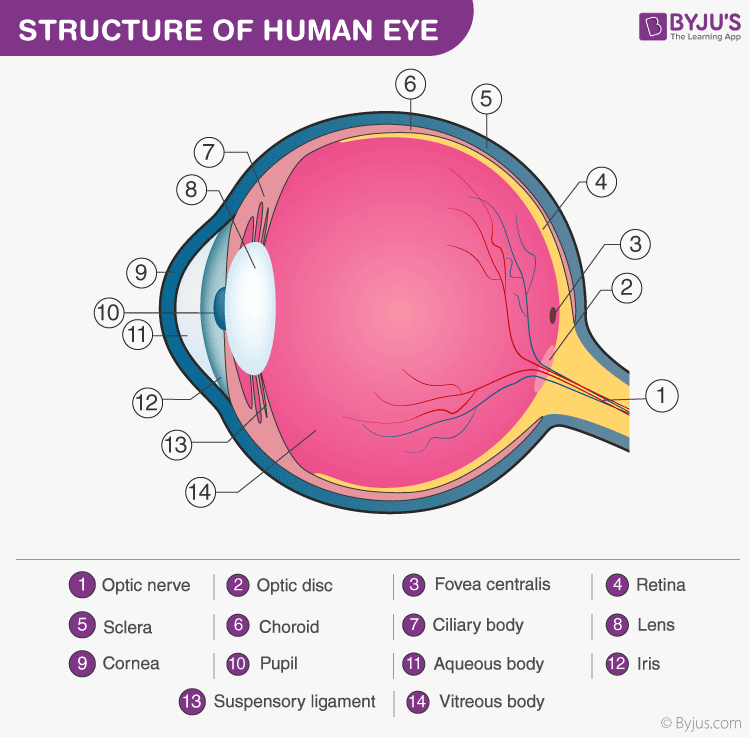
Structure and Functions of Human Eye with labelled Diagram - BYJUS The internal components of the eye include: Lens Retina Aqueous humour Optic nerve Vitreous humour Test your knowledge on Structure Of Eye Put your understanding of this concept to test by answering a few MCQs. Click 'Start Quiz' to begin! Select the correct answer and click on the "Finish" button Check your score and answers at the end of the quiz
Eye labeling diagram
eye diagram with labelling eye diagram with labelling Label the parts of the eye:. Digestive system label worksheet labeling human anatomy diagram parts labels worksheets quiz body blank overall cow systems printable practice head. Eye whyfiles parts why psychology study guide destroyed amd nih macula finest damaged vision institute graphic national site Labelled Diagram of Human Eye, Explanation and Function - VEDANTU The human eye is a part of the sensory nervous system. Labeled Diagram of Human Eye The eyes of all mammals consist of a non-image-forming photosensitive ganglion within the retina which receives light, adjusts the dimensions of the pupil, regulates the availability of melatonin hormones, and also entertains the body clock. Module 1: Labeled Diagram of the Eye - Pinterest Module 1: Labeled Diagram of the Eye Itinerary Template, Label Templates, Microsoft Word. caprockeyedoc. Wittman Vision. 3k followers. More information.
Eye labeling diagram. Module 1: Labeled Diagram of the Eye - Pinterest Module 1: Labeled Diagram of the Eye Itinerary Template, Label Templates, Microsoft Word. caprockeyedoc. Wittman Vision. 3k followers. More information. PDF Eye Anatomy Handout - National Institutes of Health Eye Anatomy Handout Author: National Eye Institute , National Eye Health Education Program Subject: Diabetes and Healthy Eyes Toolkit and Website Keywords: Eye anatomy, eye diagram, cornea, iris, lens, macula, optic nerve, pupil, retina, vitrous gel, diabetic eye disease. Created Date: 6/27/2012 11:57:40 AM Untitled Document [science.jburroughs.org] Label this diagram as you learn the name of each structure. Start this exercise by labeling your diagram of the human eye. The structures to be labeled are identified by the numbers on the diagram. Click on these numbers to learn the names of the structures. The name and additional information about the structure selected will appear in this frame. Eye Diagram: Label Quiz - PurposeGames.com This is an online quiz called Eye Diagram: Label. Points. 0. 0. to score the 11 points available. Tongue Diagram: Label 4p.
Label the Eye - The Biology Corner Label the Eye. Shannan Muskopf December 30, 2019. This worksheet shows an image of the eye with structures numbered. Students practice labeling the eye or teachers can print this to use as an assessment. There are two versions on the google doc and pdf file, one where the word bank is included and another with no word bank for differentiation. Block Diagram | Complete Guide with Examples - Edraw - Edrawsoft Dec 08, 2021 · The main goal of drawing a block diagram is to give an overview of the workflow that could be expected from the system post its completion. With the clear illustration, it becomes easy for the engineers to assess the smooth functioning of the process and to identify the existing elements (or the missing ones) that might obstruct, hinder, or unnecessarily delay the output. Labeled Eye Diagram | Science Trends What you want to interpret as a major part of the human eye is somewhat up to the individual, but in general there are seven parts of the human eye: the cornea, the pupil, the iris, the lens, the vitreous humor, the retina, and the sclera. Let's take a closer look at each of these components individually. The Cornea Labeled eye diagram - Pinterest Also labeled eye diagram and anatomy of eye and human eye structure for better ... Human eye diagram and functions with diagram of human eye with labelling.
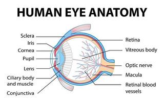
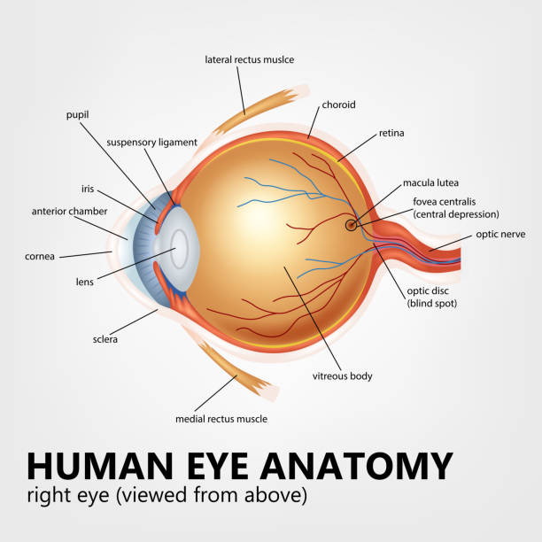
Art- labeling Activity Flashcards | Quizlet 105 terms · , , , , , , , , , , , , , , , , , , , , , , , pseudostratified columnar epithelium, simple cuboidal epithelium, Stratified squamous epithelium ... Asus ROG Zephyrus G15 laptop review: Eye-catcher 31/01/2021 · We review the Asus ROG Zephyrus G15 in the configuration with the Ryzen 9 5900HS CPU, the GeForce RTX 3080 laptop GPU, and a QHD panel. PDF Parts of the Eye - National Institutes of Health Eye Diagram Handout Author: National Eye Health Education Program of the National Eye Institute, National Institutes of Health Subject: Handout illustrating parts of the eye Keywords: parts of the eye, eye diagram, vitreous gel, iris, cornea, pupil, lens, optic nerve, macula, retina Created Date: 12/16/2011 12:39:09 PM Eye Anatomy: Parts of the Eye and How We See Behind the anterior chamber is the eye's iris (the colored part of the eye) and the dark hole in the middle called the pupil. Muscles in the iris dilate (widen) or constrict (narrow) the pupil to control the amount of light reaching the back of the eye. Directly behind the pupil sits the lens. The lens focuses light toward the back of the eye.
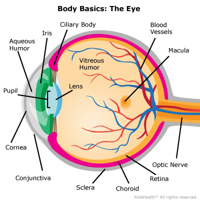
Your Eyes (for Kids) - Nemours KidsHealth
What Does the Eye Look Like? - Diagram of the Eye | Harvard Eye Associates Vitreous Gel: A thick, transparent liquid that fills the center of the eye. It is mostly water and gives the eye its form and shape. Our eyes are vital for seeing the world around us. Keep them healthy by maintaining regular vision exams. Contact Harvard Eye Associates at 949-951-2020 or harvardeye.com to schedule an appointment today.

3d Image Render Of Diagram Of Eye Anatomy With Label For ...
Label Parts of the Human Eye - University of Dayton Parts of the Eye. Select the correct label for each part of the eye. The image is taken from above the left eye. Click on the Score button to see how you did. Incorrect answers will be marked in red. ...

Correctly Label the Eye Diagram Quiz
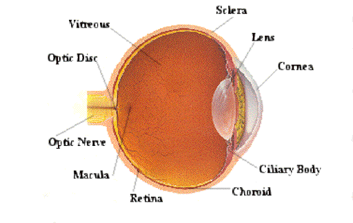
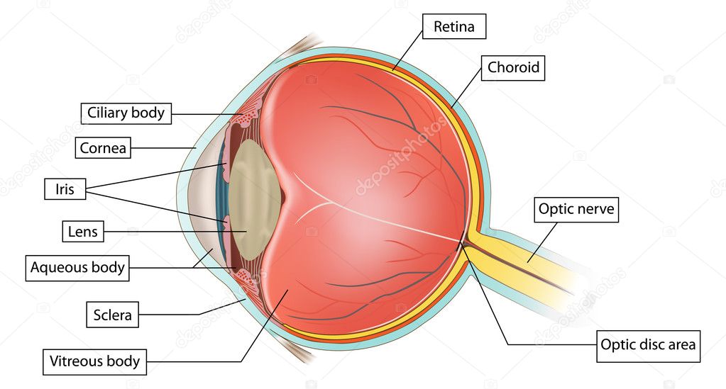
The Human Eye (Eyeball) Diagram, Parts and Pictures Diagram of the different layers of the eyeball. Outer Layer. The outer fibrous layer maintains the shape of the eyeball and protects more fragile internal structure. This layer is made up of the sclera and cornea. The sclera is the firm opaque outer part of the eye commonly referred to as the "whites" of the eye. It covers most of the outer ...

Labeled Eye Diagram | Human eye diagram, Eye anatomy, Diagram ...
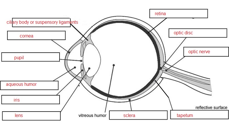
Eye Diagram With Labels and detailed description - BYJUS A brief description of the eye along with a well-labelled diagram is given below for reference. Well-Labelled Diagram of Eye The anterior chamber of the eye is the space between the cornea and the iris and is filled with a lubricating fluid, aqueous humour. The vascular layer of the eye, known as the choroid contains the connective tissue.
/GettyImages-695204442-b9320f82932c49bcac765167b95f4af6.jpg)
Structure and Function of the Human Eye
The Water Cycle Song - YouTube This is a weird song that we saw in geography.Thanks Mr leach and Mr Davies!!!!!

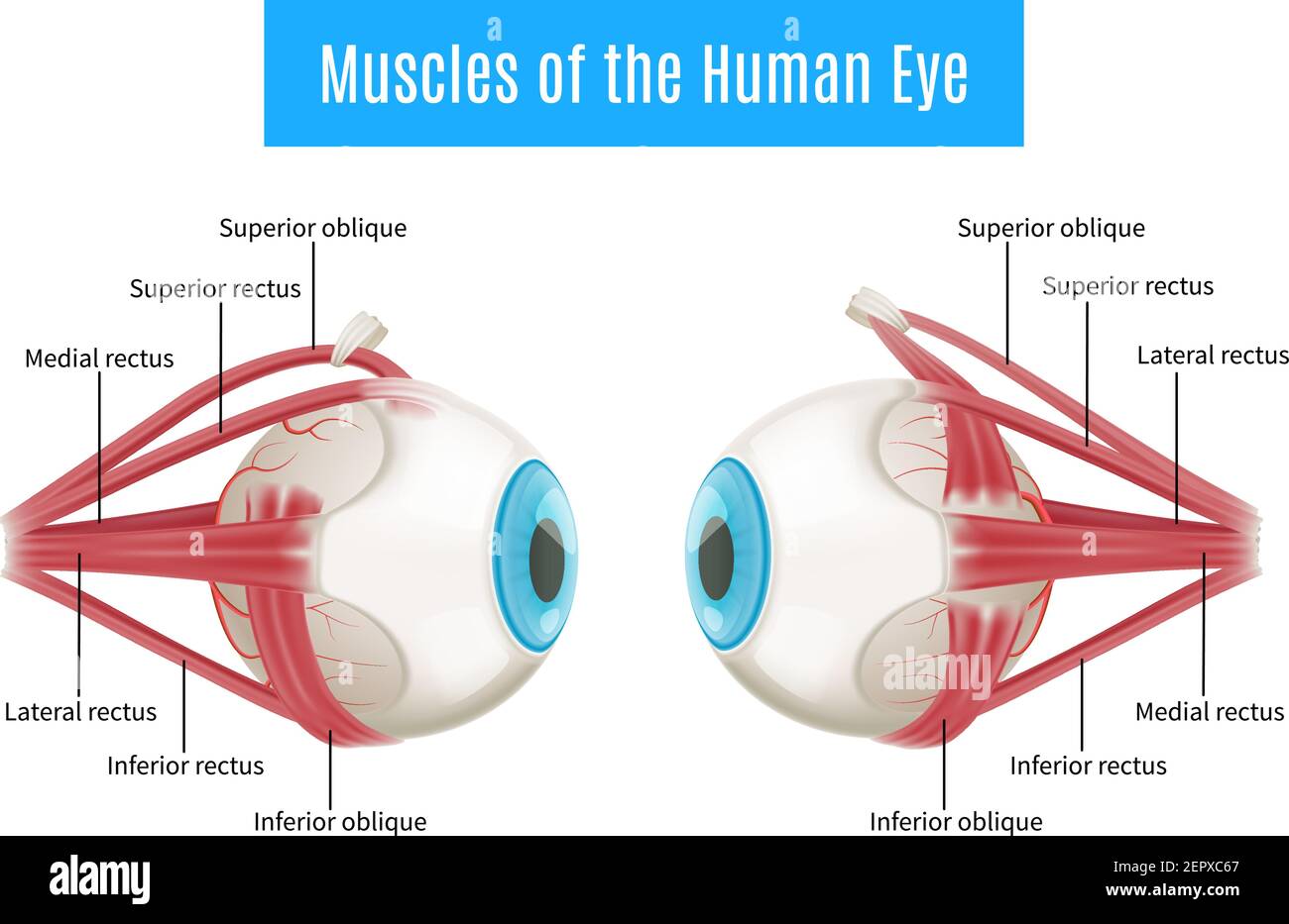
Alila Medical Media | Muscles of the eye, labeled diagram ...
Eye Anatomy: 16 Parts of the Eye & Their Functions - Vision Center It is located underneath the white part of the eye (the sclera) and is composed of three parts: The iris Ciliary body Choroid These structures control some eye functions, such as adapting to varying levels of light or object distances. If any of the structures get inflamed, the resulting condition is referred to as uveitis. 7. Choroid
WEB QUEST - Slide 6
Microscope Parts and Functions With Labeled Diagram and ... Diagram/Parts/Functions of a Compound Microscope. Beginner Microscope Experiments. Microscope Slides Preparations-Styles and Techniques. Prepared Microscope Slides - Benefits and Recommendations. See also: Dissecting Stereo Microscope Parts and Functions Stereo Microscope Vs Compound Microscope. Check out this Microscope Quiz to test your knowledge
Eye anatomy 3d diagram infographics layout showing human eyes ...
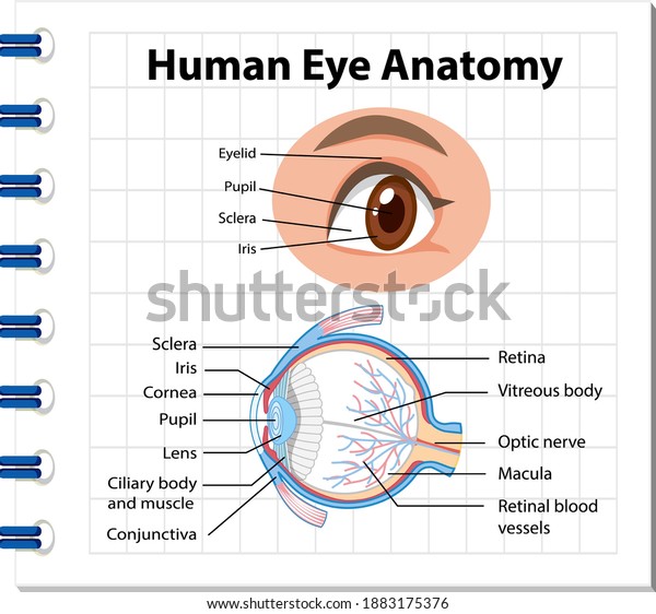
Anatomy of the Human Eye - News Medical Eyes are one of the most important organs of the body. A healthy pair of eyes means a clear vision, which plays a major role in day-to-day life and quality ...

IGCSE Label the Eye Diagram | Quizlet
Labeled Eye Diagram - Pinterest Labeled Eye Diagram. Find this Pin and more on Legume by Nicole Young. Eye Anatomy Diagram. Human Eye Diagram. Diagram Of The Eye. Human Anatomy Drawing. Human Anatomy And Physiology. Anatomy Organs. Brain Anatomy.
Eye Diagram Vector Art, Icons, and Graphics for Free Download
7 Examples of Visual Communication - Simplicable 07/09/2017 · A definition of visual communication with examples. Visual communication is the visual expression of emotion, data, information and knowledge. The term can be applied to anything visual including art, architecture, cities, products, user interfaces, media, publications, advertising, entertainment, performing art and fashion.The following are illustrative examples …
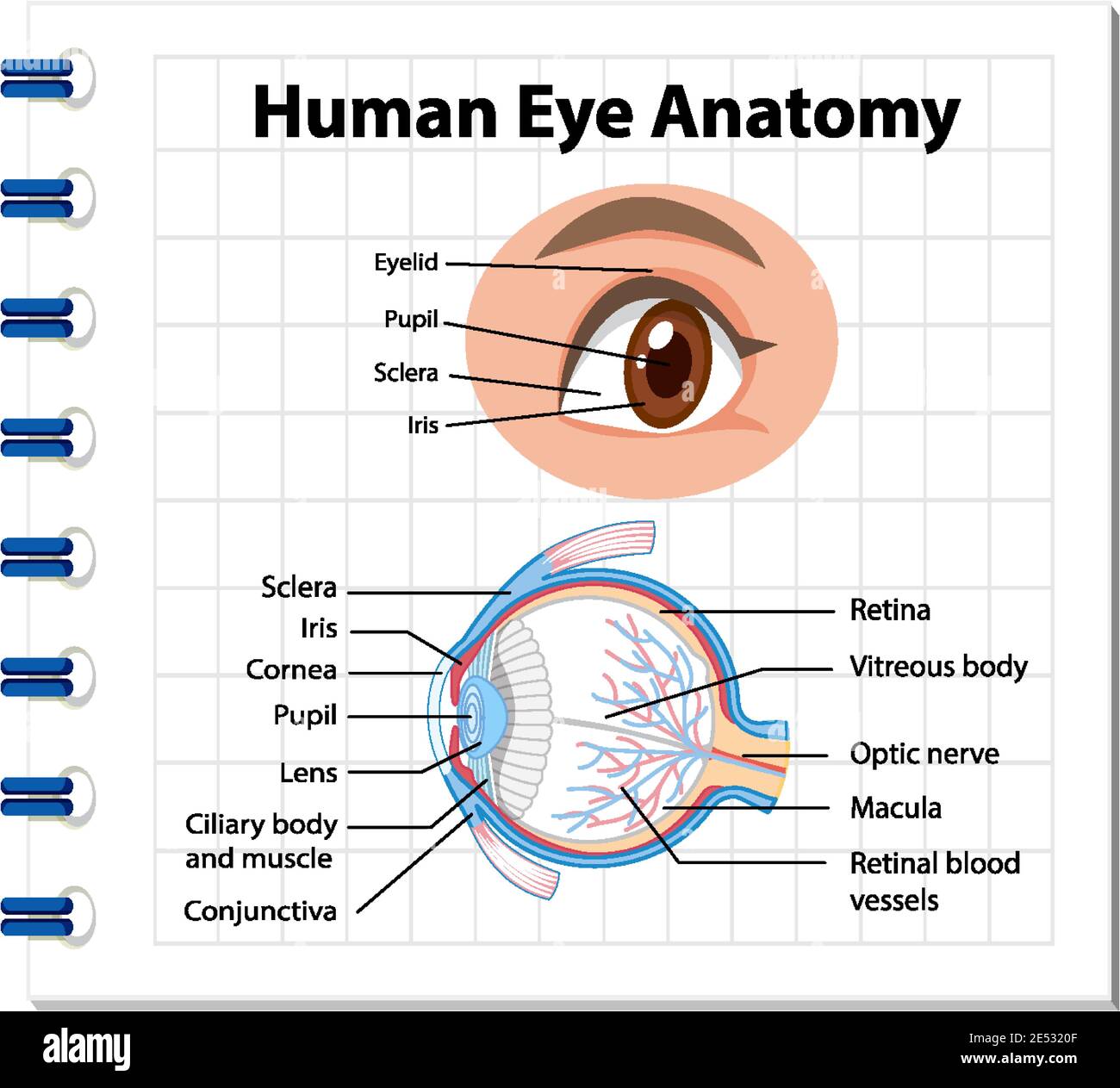
Diagram of human eye anatomy with label illustration Stock ...
Labelling the eye - Science Learning Hub In this interactive, you can label parts of the human eye. Use your mouse or finger to hover over a box to highlight the part to be named. Drag and drop the text labels onto the boxes next to the eye diagram If you want to redo an answer, click on the box and the answer will go back to the top so you can move it to another box.
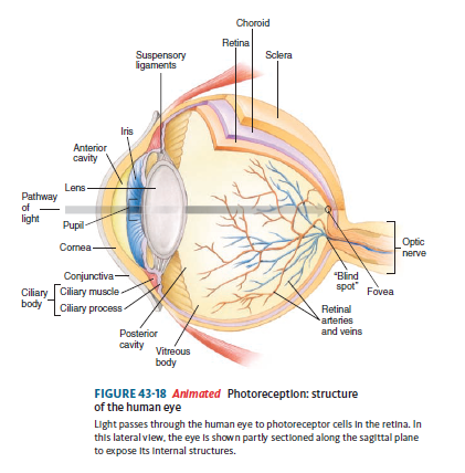
Solved: Label the diagram. Refer to Figure 43-18 to check ...
Human body organs diagram Stock Photos and Images - Alamy RFT5FEWA – Medical diagram showing the circulatory system of the human body, in particular the venous and arterial systems. RF 2CD0P4N – Human Digestive System Woman Anatomy Diagram RF 2J9168E – Flat human body posters with internal organs muscular skeletal resperatory circulatory nervous systems isolated vector illustration
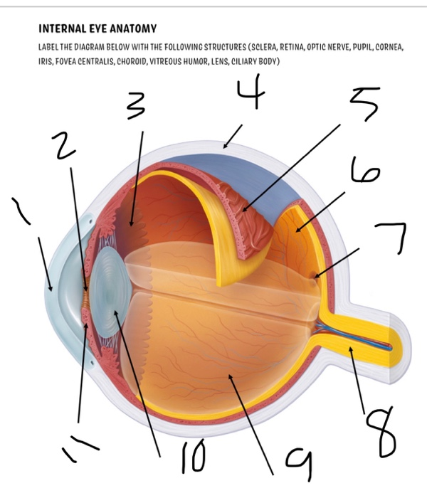
Solved INTERNAL EYE ANATOMY LABEL THE DIAGRAM BELOW WITH THE ...
Eye Diagram Quiz - ProProfs Quiz Try this amazing Eye Diagram Quiz quiz which has been attempted 5185 times by avid quiz takers. Also explore over 77 similar quizzes in this category. Take Quizzes. Animal; Nutrition; ... Quiz: Label The Parts Of The Eye. People say that the eyes are the windows to a person's soul. In the class today, we covered parts of the eye, and what ...

Diagram of Human Eye Anatomy with Label Stock Vector ...
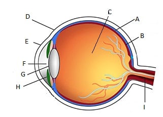
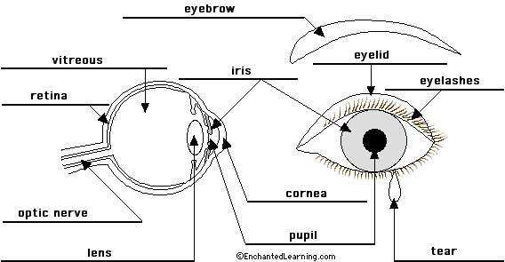
Label Eye Printout - EnchantedLearning.com Label the Eye Diagram. Human Anatomy. Read the definitions, then label the eye anatomy diagram below. Cornea - the clear, dome-shaped tissue covering the front of the eye. Iris - the colored part of the eye - it controls the amount of light that enters the eye by changing the size of the pupil. Lens - a crystalline structure located just behind ...

Anatomy of the Human Eye
The Eye Diagram: What is it and why is it used? The eye diagram is used primarily to look at digital signals for the purpose of recognizing the effects of distortion and finding its source. To demonstrate using a Tektronix MDO3104 oscilloscope, we connect the AFG output on the back panel to an analog input channel on the front panel and press AFG so a sine wave displays.
Eye anatomy Vector Art Stock Images | Depositphotos
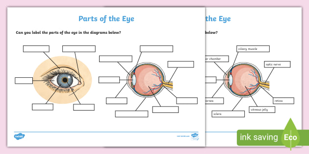
Label the Eye Worksheet - Teacher-Made Learning Resources - Twinkl In this resource, you'll find a 2-page PDF that is easy to download, print out, and use immediately with your class. The first page is a labelling exercise with two diagrams of the human eye. One is a view from the outside, and the other is a more detailed cross-section. Challenge learners to label the parts of the eye diagram. On the second page, you'll find a set of answers showing ...
Eye Anatomy Trivia Test! Quiz - ProProfs Quiz
Anatomy of the Eye Diagrams for Coloring/Labeling, with Reference and ... The core eye anatomy diagram, designed as the labeling exercise, has a fully colored and labeled reference chart to go with it. In case you need a little refresher before going over your lesson, or want something for your slightly older children to read, we have added a simply worded, but terminologically accurate summary, describing the ...

Diagram Of Eye For Kids The Eye Diagram For Kids Nurse ...
5th grade science games, interactive quiz games - Ecosystem For … Human Organs labeling game. Play here >>> Atoms & elements. Jungle girl board game on facts related to atoms and elements - 5th grade quiz. Play here >>> Cells and functions. Learn the parts of a cell through an online pirate game for 5th grade children. Play here >>> Earth movements. 5th grade science quiz on Earth movements; erosion, landslides, mudlow etc. Play here >>> …

Human Eye Ball Anatomy & Physiology Diagram
eye labeling Diagram | Quizlet It is the first structure to refract (bend) light that enters the eye. sclera Tough white out covering of the eyeball choroid Middle layer of the eye (between the retina and the sclera) that contains the blood vessels that nourish the eye and cornea iris colored layer that dilates and constricts to allow in more or less light ciliary body
Eye Anatomy Diagram - EnchantedLearning.com
Labelled diagram - Wordwall Labelled diagram Drag and drop the pins to their correct place on the image. ... The Eye. by Pamela209. Labelled diagram. ... Labeling Microscope. by Jessicamoser ...

Diagram human eye anatomy with label Royalty Free Vector
Anatomy of the eye: Quizzes and diagrams | Kenhub Take a look at the diagram of the eyeball above. Here you can see all of the main structures in this area. Spend some time reviewing the name and location of each one, then try to label the eye yourself - without peeking! - using the eye diagram (blank) below. Unlabeled diagram of the eye
Human Eye - Definition, Structure, Function, Parts, Diagram
Beef Cuts Chart and Diagram, with Photos, Names, Recipes, and ... Jul 08, 2022 · Ingredients Beef Cuts Chart and Diagram, with Photos, Names, Recipes, and More. Learn all about the most popular beef cuts from our chart, diagram and write up, including popular and alternative names, where the cuts come from on the cow, preferred outdoor cooking methods, their costs relative to each other, and a fantastic recipe for each cut of beef that we’ve taken from around the web.
4,189 Eye Diagram Stock Photos, Pictures & Royalty-Free ...
Eye labeling Diagram | Quizlet Iris a ring of muscle tissue that forms the colored portion of the eye around the pupil and controls the size of the pupil opening Cornea The clear tissue that covers the front of the eye Posterior Compartment filled with vitreous humor Pupil opening in the center of the iris Susponsory Ligament Allows the eye to move up and down

The Structure of the Eye Picture Hotspots (teacher made)
Cow's Eye Dissection - Eye diagram - Exploratorium A clear fluid that helps the cornea keep its rounded shape. The pupil is the dark circle in the center of your iris. It's a hole that lets light into the inner eye. Your pupil is round. A cow's pupil is oval. A tough, clear covering over the iris and the pupil that helps protect the eye. Light bends as it passes through the cornea.

Free eye anatomy - Vector Art
Human eye diagram, Eye anatomy, Diagram of the ... - Pinterest Human Eye Diagram Labeled - Health, Medicine and Anatomy Reference Pictures Eye Anatomy Diagram,. Visit. Save. From. healthfavo.com ...

L2 BTEC Animal Care: Label the Mammal Eye Diagram | Teaching ...
Diagram of the Eye - Lions Eye Institute Instructions Click the parts of the eye to see a description for each. Hover the diagram to zoom. Need any help? If you would like to know more about us, or want to make an appointment, please don't hesitate to get in touch. (08) 9381 0777 carecentre@lei.org.au Request an appointment Customer Care Centre (08) 9381 0777

Diagram human eye anatomy with label Royalty Free Vector
Module 1: Labeled Diagram of the Eye - Pinterest Module 1: Labeled Diagram of the Eye Itinerary Template, Label Templates, Microsoft Word. caprockeyedoc. Wittman Vision. 3k followers. More information.

4,189 Eye Diagram Stock Photos, Pictures & Royalty-Free ...
Labelled Diagram of Human Eye, Explanation and Function - VEDANTU The human eye is a part of the sensory nervous system. Labeled Diagram of Human Eye The eyes of all mammals consist of a non-image-forming photosensitive ganglion within the retina which receives light, adjusts the dimensions of the pupil, regulates the availability of melatonin hormones, and also entertains the body clock.

Human Eye & Ear Diagram Labeling Worksheet - Science
eye diagram with labelling eye diagram with labelling Label the parts of the eye:. Digestive system label worksheet labeling human anatomy diagram parts labels worksheets quiz body blank overall cow systems printable practice head. Eye whyfiles parts why psychology study guide destroyed amd nih macula finest damaged vision institute graphic national site

Structures Of The Human Eye Labeled art print poster
FREE! - Label the Eye Worksheet – Teacher-Made Learning Resources

Eye Anatomy | Human Anatomy Quiz - Quizizz
Cow Eye Dissection

Eye diagram to be labeled worksheet
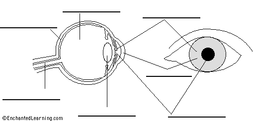
Label Eye Printout - EnchantedLearning.com

eye labeling Diagram | Quizlet

Module 1: Labeled Diagram of the Eye | Diagram of the eye ...
Vektor Stok Diagram Human Eye Anatomy Label Illustration ...

human eye | Definition, Anatomy, Diagram, Function, & Facts ...

Diagram of human eye anatomy with label illustration. | CanStock

Eye anatomy labeled diagram | Alila Medical Images
Post a Comment for "41 eye labeling diagram"