44 microscope diagram unlabeled
Electrochemiluminescence biosensor for determination of lead(II) … Aug 05, 2022 · MXene@Au as the base and Au@SiO2 as signal amplification factor were used for constructing an ultrasensitive “on–off” electrochemiluminescence (ECL) biosensor for the detection of Pb2+ in water. The use of MXene@Au composite provided a good interface environment for the loading of tris(2,2-bipyridyl)ruthenium(II) (Ru(bpy)32+) on the electrode. … Unlabeled Microscope Diagram - Cliparts.co Unlabeled Microscope Diagram 59 images of Unlabeled Microscope Diagram. You can use these free cliparts for your documents, web sites, art projects or presentations. Don't forget to link to this page for attribution!
A Study of the Microscope and its Functions With a Labeled Diagram ... Here, unlabeled microscope diagrams have been provided for your perusal, which will help you practice and test your understanding of the instrument. Types of Microscopes Depending on the source of illumination, microscopes can be divided into two categories. They are:
Microscope diagram unlabeled
Fundamental Concepts in DIC Microscopy - Life Science The basic differential interference contrast (DIC) system, first devised by Francis Smith in 1955, is a modified polarized light microscope with two Wollaston prisms added, one to the front focal plane of the condenser and a second at the rear focal plane of the objective (see Figure 1).Several years later, Georges Nomarski, a Polish-born French physicist, modified the … Biology Ch. 4 (Unlabeled Microscope) Diagram | Quizlet Start studying Biology Ch. 4 (Unlabeled Microscope). Learn vocabulary, terms, and more with flashcards, games, and other study tools. Looking at the Structure of Cells in the Microscope A light microscope. (A) Diagram showing the light path in a compound microscope. Light is focused on the specimen by lenses in the condensor. ... to an antibody used for specific recognition—the primary antibody—a stronger signal is achieved by using an unlabeled primary antibody and then detecting it with a group of labeled secondary ...
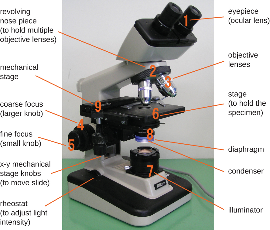
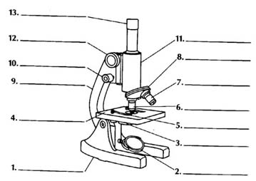
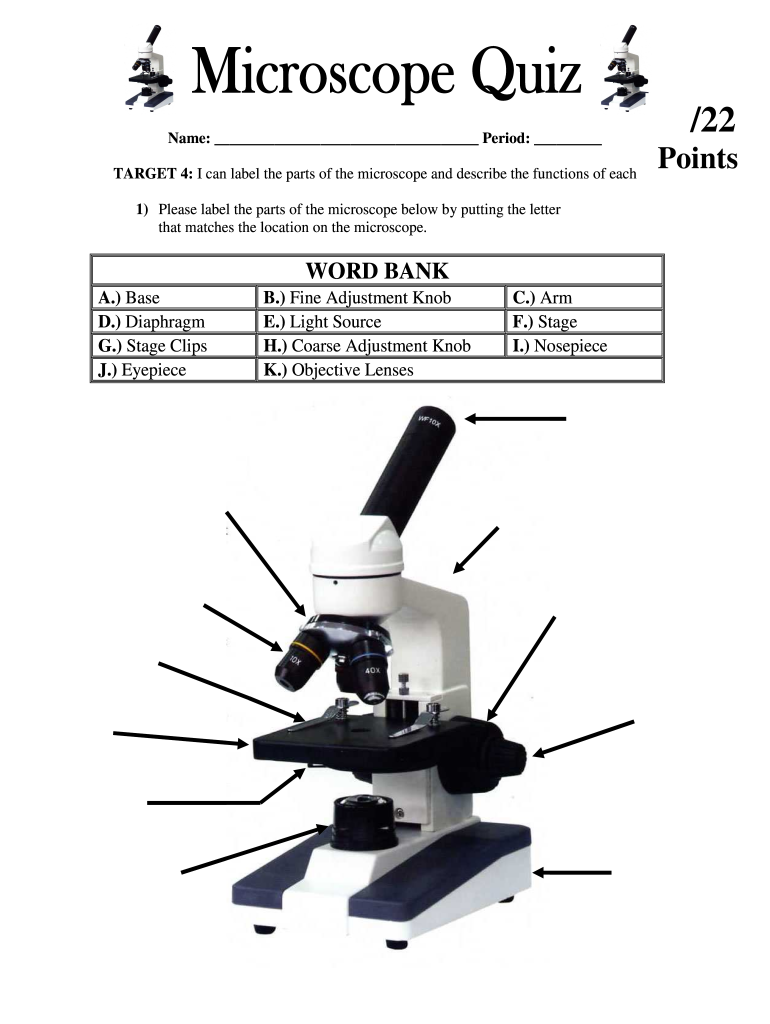
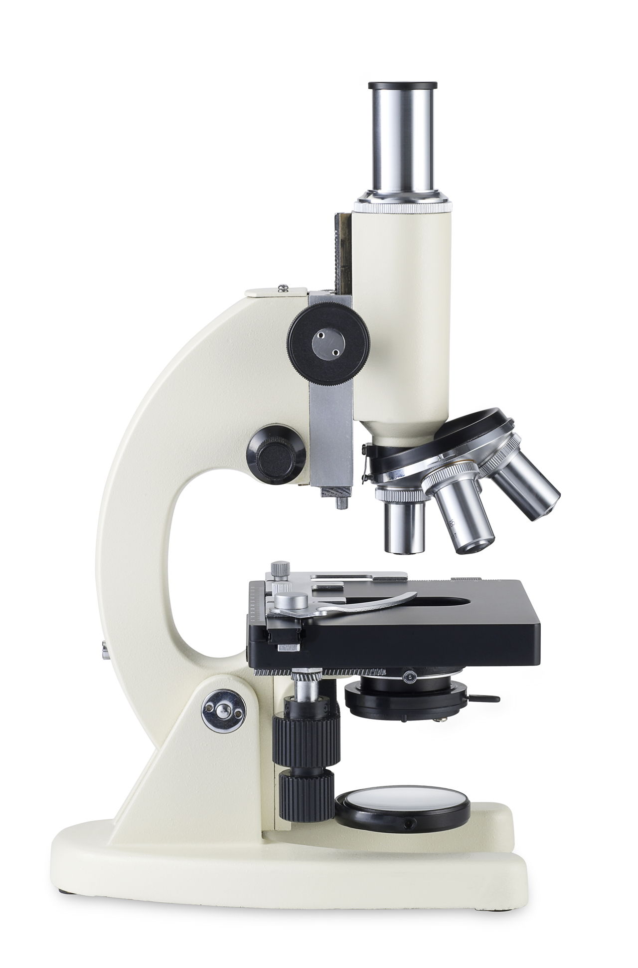
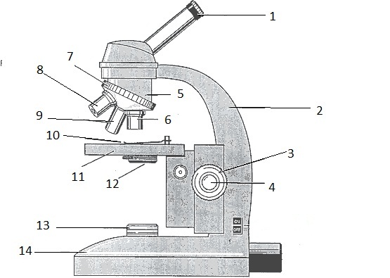
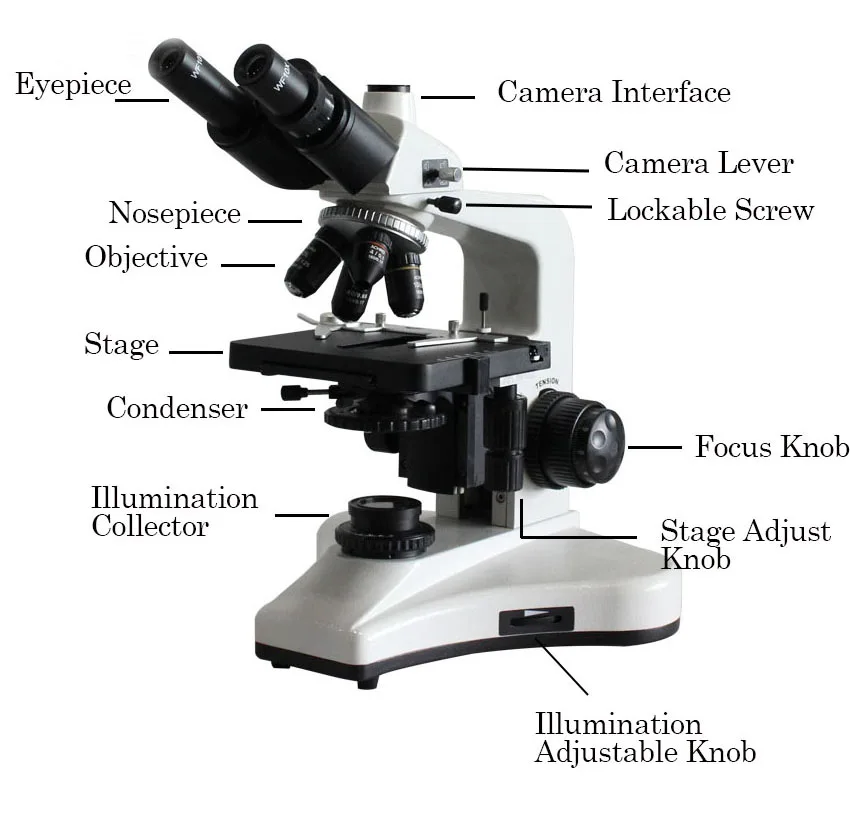
Microscope diagram unlabeled. Junqueira's Basic Histology Text and Atlas, 14th Edition There is shortage of references in higher teaching institutions especially in newly opened institutions engaged in training of various Veterinary professionals in the country. Microscope Diagram Labeled, Unlabeled and Blank - Pinterest Description Worksheet identifying the parts of the compound light microscope. Answer key: 1. Body tube 2. Revolving nosepiece 3. Low power objective 4. Medium power objective 5. High power objective 6. Stage clips 7. Diaphragm 8. Light source 9. Eyepiece 10. Arm 11. Stage 12. Coarse adjustment knob 13. Fine adjustment knob 14. Base S Antigen-B Cell Receptor Complexes Associate with Intracellular … Nov 06, 2015 · Introduction. T lymphocyte activation is driven by the recognition of complexes of antigen-derived peptide and MHC molecules on the surface of an antigen-presenting cell (APC). 2 Cytotoxic CD8-positive T cells recognize peptide-MHC class I complexes and generally kill the APC, whereas helper CD4-positive T cells recognize peptide-MHC class II complexes and … Microscope Parts and Functions A standard microscope has three, four, or five objective lenses that range in power from 4X to 100X. When focusing the microscope, be careful that the objective lens doesn't touch the slide, as it could break the slide and destroy the specimen. Specimen or slide: The specimen is the object being examined.
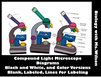
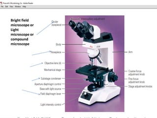
Microscope Diagram Labeled, Unlabeled and Blank | Parts of a Microscope ... Microscope Diagram Labeled, Unlabeled and Blank | Parts of a Microscope Print a microscope diagram, microscope worksheet, or practice microscope quiz in order to learn all the parts of a microscope. Tim's Printables 38k followers More information Microscope Diagram Unlabeled Find this Pin and more on Chemistry by Ginger Dwyer. Labeled Microscope and Basics of Life Diagram | Quizlet PLAY. A microscope is an instrument widely to magnify and resolve the image of an object that is otherwise invisible to naked eye. For resolving the details of objects, which otherwise cannot be achieved by naked eye, a microscope is used. This set of flash cards will help the student to identify the different parts and function of the microscope. Molecular mechanism for the synchronized electrostatic ... - Nature Aug 06, 2022 · Here, the authors report that α-synuclein phase-separates into liquid condensates with positively charged polypeptides such as Tau. The condensates undergo different maturation processes ... 5 Optical Mineralogy – Mineralogy - OpenGeology Most optical mineralogy today involves specially prepared thin sections (0.03-mm-thick specimens of minerals or rocks mounted on glass slides).Video 1 (linked in Box 5-2) explains how we make thin sections, and Figure 5.1, the opening figure in this chapter, shows an example. Figure 5.4 above shows a microscope view of a thin section that contains several minerals …
Label the microscope — Science Learning Hub Drag and drop the text labels onto the microscope diagram. If you want to redo an answer, click on the box and the answer will go back to the top so you can move it to another box. If you want to check your answers, use the Reset incorrect button. This will reset incorrect answers only. parts of a microscope diagram Microscope Illustration Parts - Micropedia. 18 Images about Microscope Illustration Parts - Micropedia : Microscope Diagram to Print, Microscope Diagram Labeled, Unlabeled and Blank | Parts of a Microscope and also Microscope Illustration Parts - Micropedia. Microscope Illustration Parts - Micropedia microspedia.blogspot.com Login - NotebookingPages.com Microscope pages; Notebooking templates; Blank grid paper; ... labeled and unlabeled in both portrait and landscape layouts) Table pages for element data and details; Individual element data pages ... Bird Study Pages (cover pages, Bird Log pages, Parts of a Bird diagram pages, plus pages for the study of eyes, ears, beaks, feet, facts ... Compound Microscope Parts - Labeled Diagram and their Functions Labeled diagram of a compound microscope Major structural parts of a compound microscope There are three major structural parts of a compound microscope. The head includes the upper part of the microscope, which houses the most critical optical components, and the eyepiece tube of the microscope.
Microscope Png Transparent, Biology Microscope Science ...
PDF Parts of a Microscope Printables - Homeschool Creations Microscopes help us see small organisms, or specimens. The various lenses on a microscope make the specimen appear much larger, or magnified, much more than the human eye can observe on its own. •Which part of the microscope do you look through to see a specimen? the eyepiece

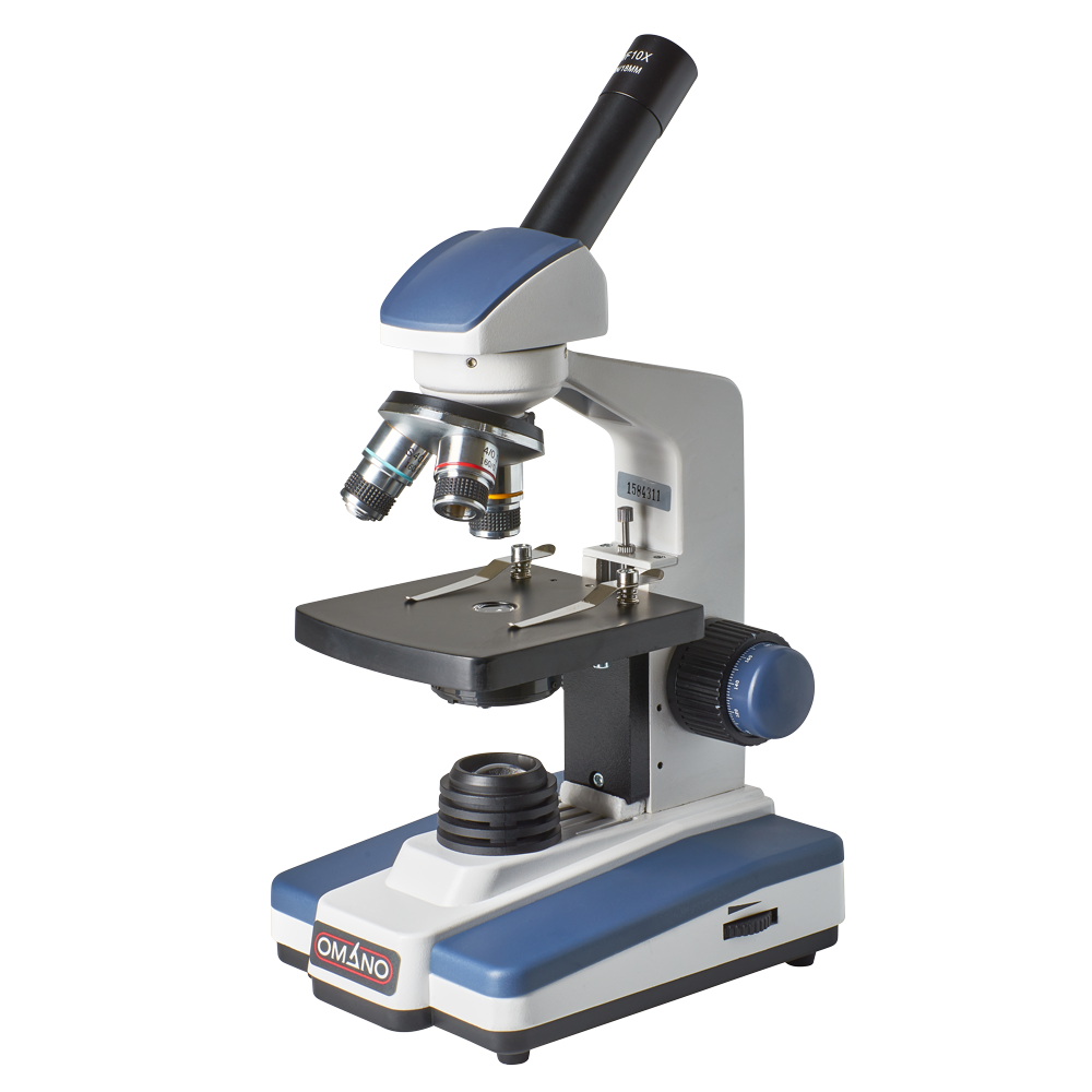
OMAX Microscope AmScope Student Stereo Binocular Microscope ...
Top 10 microscope ideas and inspiration - pinterest.com The Barska 40x - 2000x Compound Binocular Microscope delivers impressive detail and clarity with both a 10x and 20x wide field eyepiece, and a set of 4x, 10x, 40x, and 100x objective lenses . A 20 watt light with adjustable brightness allows the user to set the brightness setting while viewing a specimen.
2.3 Instruments of Microscopy – Microbiology: Canadian Edition
Microscope Labeling Game - PurposeGames.com About this Quiz. This is an online quiz called Microscope Labeling Game. There is a printable worksheet available for download here so you can take the quiz with pen and paper. This quiz has tags. Click on the tags below to find other quizzes on the same subject. Science.

Amazon.com: OMAX 40X-800X Digital Monocular Compound ...
Collection of Unlabeled Microscope Diagram (55) - Clipart Library Download Unlabeled Microscope Diagram and use any clip art,coloring,png graphics in your website, document or presentation. Collection of Unlabeled Microscope Diagram (55) drawing simple microscope large intestine and small intestine diagram stomach diagram amoeba diagram without labeled

Microscope Diagram Labeled, Unlabeled and Blank | Parts of a ...
Microscope Diagram Teaching Resources | Teachers Pay Teachers Microscope Crossword with Diagram | Printable & Distance Learning Options by Tangstar Science 5.0 (43) $2.99 Zip Google Apps™ Updated for distance learning. This resource comes in three versions (with full answer keys): 1) Editable Word Document 2) PDF and 3) Google Slide where students can type directly into the crossword boxes.
blank diagram of a compound light microscope - Clip Art Library
Atlas of Subcellular RNA Localization Revealed by APEX-Seq Jul 11, 2019 · To test APEX-catalyzed RNA biotinylation in living cells, we generated HEK cells stably expressing APEX2 in the cytosol. We labeled the cells with BP and H 2 O 2 for 1 min, extracted total RNA, and analyzed the RNA by streptavidin dot blot. Figure 1B shows that RNA biotinylation is abolished upon omission of BP or H 2 O 2 or following treatment with RNase.

Kit Mikroskop Biologis untuk Lab Klinis Dokter Hewan Kampus --- SWIFT 40X-2500X LED Pro Mikroskop Senyawa Teropong + Kamera + Slide
microscope label worksheet 17 Best Images Of Blank Microscope Worksheet - Blank Microscope Diagram microscope diagram blank worksheet labeled labeling parts compound worksheeto heart light unlabeled via quiz FREE Parts Of A Microscope Label Worksheet homeschoolgiveaways.com Microscope-diagram-unlabeled.jpg (927×1200).

AmScope 40X-2000X Professional Kohler Binocular Compound Microscope (B660B)
Parts of a Microscope Quiz - Sporcle Top Contributed Quizzes in Science. 1. Click the Laboratory Equipment. 2. Click a Seal. 3. Hidden Odd One Out: Math. 4. Clueless Letter Lines: Solar System Moons.

Types of Microscopes Unit 4 Biology. - ppt download
microscope practice worksheet Microscope-Diagram-Unlabeled.jpg (927×1200) | Science Diagrams, Science microscope diagram unlabeled light blank parts biology science labeled lab sketch worksheet labels timvandevall quiz chemistry fill blanks worksheets labs Label The Parts Of The Light Microscope youcansharethe.blogspot.com microscope parts light label

BIO: The Microscope Diagram | Quizlet
20 Color the Microscope Parts Worksheet | Worksheet From Home Microscope Parts Diagram PDF Science Printables color the microscope parts worksheet answers, color the microscope parts worksheet, via: timvandevall.com. Numbering Worksheets for Kids. Kids are usually introduced to this topic matter during their math education. The main reason behind this is that learning math can be done with the worksheets.

Microscope Diagram - Free Printable Tests and Worksheets ...
Microscope Diagram Labeled, Unlabeled and Blank - Pinterest Description Worksheet identifying the parts of the compound light microscope. Answer key: 1. Body tube 2. Revolving nosepiece 3. Low power objective 4. Medium power objective 5. High power objective 6. Stage clips 7. Diaphragm 8. Light source 9. Eyepiece 10. Arm 11. Stage 12. Coarse adjustment knob 13. Fine adjustment knob 14. Base S

7 Screen capture of a unlabeled, digitized histology image ...
A phosphate starvation response-centered network Oct 28, 2021 · (A) The schematic diagram shows the position of P1BS in OsRAM1, OsWRI5A, OsPT11 and OsAMT3;1 promoters. (B) EMSA assay showing specific binding of OsPHR2 to the P1BS element. The unlabeled mP1BS (GNAGAGNC) containing DNA fragment (10-fold, 50-fold, 100-fold) was used as a competitor.
Microscope Diagram Teaching Resources | Teachers Pay Teachers
Looking at the Structure of Cells in the Microscope A light microscope. (A) Diagram showing the light path in a compound microscope. Light is focused on the specimen by lenses in the condensor. ... to an antibody used for specific recognition—the primary antibody—a stronger signal is achieved by using an unlabeled primary antibody and then detecting it with a group of labeled secondary ...

Microscope Use Lab Purpose: To learn the parts and how to use ...
Biology Ch. 4 (Unlabeled Microscope) Diagram | Quizlet Start studying Biology Ch. 4 (Unlabeled Microscope). Learn vocabulary, terms, and more with flashcards, games, and other study tools.
Microscope Fill In The Blank - Fill Online, Printable ...
Fundamental Concepts in DIC Microscopy - Life Science The basic differential interference contrast (DIC) system, first devised by Francis Smith in 1955, is a modified polarized light microscope with two Wollaston prisms added, one to the front focal plane of the condenser and a second at the rear focal plane of the objective (see Figure 1).Several years later, Georges Nomarski, a Polish-born French physicist, modified the …

Perhatikan gambar mikroskop di bawah ini! Bagia...

Compound Light Microscope Diagram | Quizlet
Understanding the Compound Microscope Parts and its Functions ...
Microscopy

VEE GEE 1323PHi VanGuard Compound Microscope User Manual ...
Human Anatomy
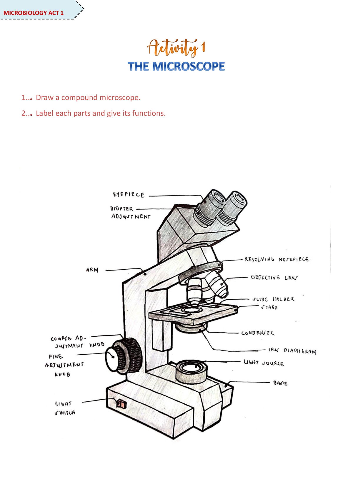
Microscope Activity - MICROBIOLOGY - 1... Draw a compound ...
Parts of a Microscope | Biology - Quizizz

Learning by Questions

Microscope Diagram Labeled, Unlabeled and Blank | Parts of a ...

Labeling the parts of a dissecting microscope Quiz

AmScope on Twitter: "Check out this awesome compact binocular ...

Parts of a Microscope (Pre-Assessment) | Science - Quizizz
Optical microscope diagram hi-res stock photography and ...

Label the parts of this microscope. please don't give other ...

Mekanik Ganda Platform Mobile Mikroskop 40-1600X Trinocular Biological Microscope Rumah Pendidikan Mikroskop Anak-anak

Diagrams Cell Biology GCSE (AQA) | Teaching Resources

Microscopy | biology

Microscope With Labels clip art | Microscope parts ...
Microscope Lab Learn the parts which are directly used for ...

The Compound Microscope parts & how ... | Microscope parts ...

Microscope Diagram Labeled, Unlabeled and Blank | Parts of a ...

Living Environment Course

Optical Microscopes | Traditional Microscopes | Grovers Optics
Bl-123 Series 40x~1600x Trinocular Biological Microscope ...

Microscopes | Biology - Quizizz
Microscope 40X / 100X / 400X / 1000X (Model 3000F-100-LED)
Parts of a Microscope Foldable-Unlabeled

The Neuron Motor neuron, Unlabeled Microscope Diagram ...
Post a Comment for "44 microscope diagram unlabeled"