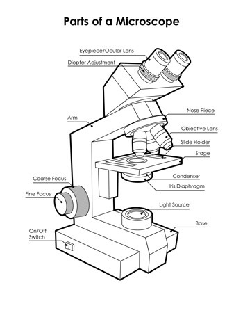
45 diagram of a microscope labeled
Lymphocytes Under Microscope with Labeled Diagram Lymphocytes Under Microscope with Labeled Diagram 10/12/2022 by anatomylearner Lymphocytes under a microscope show a round to slightly indented nucleus with clumped heterochromatin. You know these lymphocytes are agranulocytes and form the second-largest population of white blood cells. Microscope Labeling Diagram | Quizlet Coarse Focus Knob Moves the stage large distances to roughly focus the image. Fine Focus Knob Moves the stage tiny distances to slightly adjust and fine-tune the image focus. Arm Supports the body tube. Objective Lenses Focus and magnify light in differing amounts to view the specimen. Stage Clips Hold the slide in place on the stage. Nosepiece
16 Parts of a Compound Microscope: Diagrams and Video The 16 core parts of a compound microscope are: Head (Body) Arm Base Eyepiece Eyepiece tube Objective lenses Revolving Nosepiece (Turret) Rack stop Coarse adjustment knobs Fine adjustment knobs Stage Stage clips Aperture Illuminator Condenser Diaphragm Video: Parts of a compound Microscope with Diagram Explained

Diagram of a microscope labeled
Microscope Parts and Functions Most specimens are mounted on slides, flat rectangles of thin glass. The specimen is placed on the glass and a cover slip is placed over the specimen. This allows the slide to be easily inserted or removed from the microscope. It also allows the specimen to be labeled, transported, and stored without damage. Labeled Microscope and Basics of Life Diagram | Quizlet Created by. Chevron_Fickel. A microscope is an instrument widely to magnify and resolve the image of an object that is otherwise invisible to naked eye. For resolving the details of objects, which otherwise cannot be achieved by naked eye, a microscope is used. This set of flash cards will help the student to identify the different parts and ... 16 Essential Microscope Parts: Names, Functions & Labeled Diagram Microscope Parts Labeled Diagram The principle of the Microscope gives you an exact reason to use it. It works on the 3 principles. Magnification Resolving Power Numerical Aperture. Parts of Microscope Head Base Arm Eyepiece Lens Eyepiece Tube Objective Lenses Nose Piece Adjustment Knobs Stage Aperture Microscopic Illuminator Condenser Lens
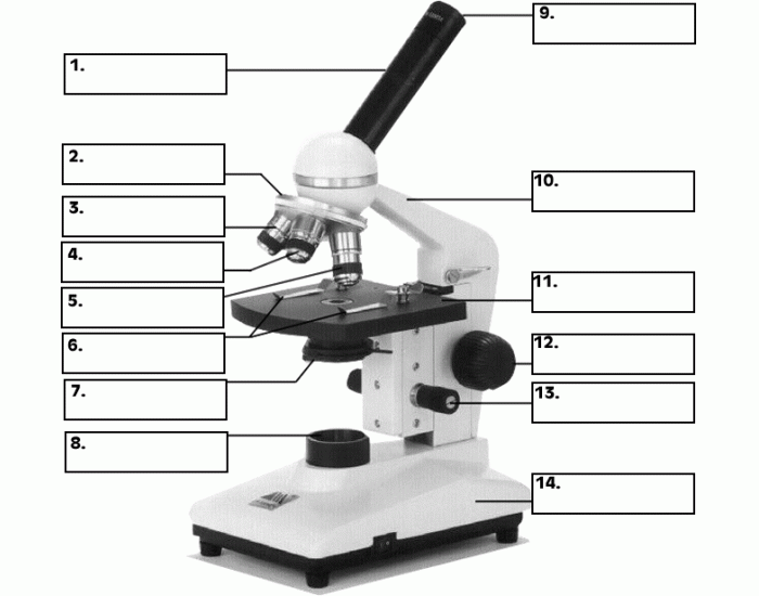
Diagram of a microscope labeled. Labelled Diagram of Compound Microscope The below mentioned article provides a labelled diagram of compound microscope. Part # 1. The Stand: The stand is made up of a heavy foot which carries a curved inclinable limb or arm bearing the body tube. The foot is generally horse shoe-shaped structure (Fig. 2) which rests on table top or any other surface on which the microscope in kept. Labeling the Parts of the Microscope | Microscope World Resources Labeling the Parts of the Microscope This activity has been designed for use in homes and schools. Each microscope layout (both blank and the version with answers) are available as PDF downloads. You can view a more in-depth review of each part of the microscope here. Download the Label the Parts of the Microscope PDF printable version here. Cardiac Muscle Under Microscope with Labeled Diagram The skeletal muscle under a microscope with labeled diagram, So, from the fine structure of cardiac muscle, I will show you the following features - Cardiac muscle cell and centered nucleus, Organelles and glycogen stored in myofibrils, Intercalated disc and different cell junctions, Sarcomere and sarcoplasm of cardiac muscle fibers, and Parts of a microscope with functions and labeled diagram - Microbe Notes Figure: Diagram of parts of a microscope There are three structural parts of the microscope i.e. head, base, and arm. Head - This is also known as the body. It carries the optical parts in the upper part of the microscope. Base - It acts as microscopes support. It also carries microscopic illuminators.
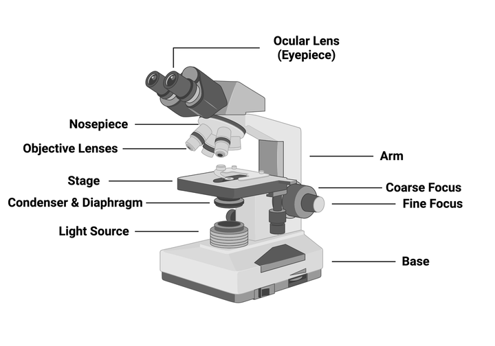
A Study of the Microscope and its Functions With a Labeled Diagram ... A Study of the Microscope and its Functions With a Labeled Diagram To better understand the structure and function of a microscope, we need to take a look at the labeled microscope diagrams of the compound and electron microscope. These diagrams clearly explain the functioning of the microscopes along with their respective parts. Compound Microscope Labeled Diagram | Quizlet Start studying Compound Microscope Labeled. Learn vocabulary, terms, and more with flashcards, games, and other study tools. Tongue Under Microscope with Labeled Diagram - AnatomyLearner Tongue Under Microscope with Labeled Diagram 19/11/2022 by anatomylearner The tongue under a microscope shows a core of crisscrossing skeletal muscle bundles and a peripheral mucous membrane. A stratified squamous epithelium covers the mucous membrane of the tongue and contains 4 types of papillae. Parts of the Microscope (Labeled Diagrams) Simple microscope labelled diagram Image created with Biorender Tube/Body Tube It serves as the connector between the eyepiece/ocular and objective lenses. Objective lenses The lenses have varying magnifying power, which typically consists of 10x, 40x, and 100x.
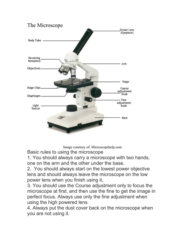
PDF Label parts of the Microscope: Answers Label parts of the Microscope: Answers Coarse Focus Fine Focus Eyepiece Arm Rack Stop Stage Clip . Created Date: 20150715115425Z ... Light microscopes - Cell structure - Edexcel - BBC Bitesize The components of a light microscope and their functions Calculating the magnification of light microscopes. The compound microscope uses two lenses to magnify the specimen: the eyepiece and an ... Microscope Types (with labeled diagrams) and Functions Simple microscope labeled diagram Simple microscope functions It is used in industrial applications like: Watchmakers to assemble watches Cloth industry to count the number of threads or fibers in a cloth Jewelers to examine the finer parts of jewelry Miniature artists to examine and build their work Also used to inspect finer details on products Label Microscope Diagram - EnchantedLearning.com Using the terms listed below, label the microscope diagram. arm - this attaches the eyepiece and body tube to the base. base - this supports the microscope. body tube - the tube that supports the eyepiece. coarse focus adjustment - a knob that makes large adjustments to the focus. diaphragm - an adjustable opening under the stage, allowing ...
Label the microscope — Science Learning Hub
Compound Microscope: Definition, Diagram, Parts, Uses, Working ... - BYJUS Compound microscope is a type of optical microscope that is used for obtaining a high-resolution image. There are more than two lenses in a compound microscope. Learn about the working principle, parts and uses of a compound microscope along with a labeled diagram here.

Types, Parts and Functions of a Microscope
Label the microscope — Science Learning Hub Label the microscope Interactive Add to collection Use this interactive to identify and label the main parts of a microscope. Drag and drop the text labels onto the microscope diagram. eye piece lens diaphragm or iris coarse focus adjustment stage base fine focus adjustment light source high-power objective Download Exercise Tweet
1.2: Microscopes - Biology LibreTexts
Simple Microscope - Diagram (Parts labelled), Principle, Formula and Uses A simple microscope consists of Optical parts Mechanical parts Labeled Diagram of simple microscope parts Optical parts The optical parts of a simple microscope include Lens Mirror Eyepiece Lens A simple microscope uses biconvex lens to magnify the image of a specimen under focus.
Lab :1
Microscope Parts, Types & Diagram | What is a Microscope? Microscope Diagram There are many illustrations available for the diagram of a light microscope. The essential parts include the head, base, arms, lenses, and lights. In diagrams, one...

Types, Parts and Functions of a Microscope
Colon Histology Slide with Labeled Diagram - AnatomyLearner In addition, the colon labeled diagram also shows the bundle of nerve fibers (not seen under the binocular microscope). Finally, the labeled diagram shows a thin layer of tunica serosa that lines with a single layer of squamous cells. The mucosa of a colon labeled diagram. Let's see the second labeled diagram of the animal colon.

label microscope diagram | Charts | Microscope, Diagram chart ...
Parts of the Microscope with Labeling (also Free Printouts) A microscope is one of the invaluable tools in the laboratory setting. It is used to observe things that cannot be seen by the naked eye. Table of Contents 1. Eyepiece 2. Body tube/Head 3. Turret/Nose piece 4. Objective lenses 5. Knobs (fine and coarse) 6. Stage and stage clips 7. Aperture 9. Condenser 10. Condenser focus knob 11. Iris diaphragm

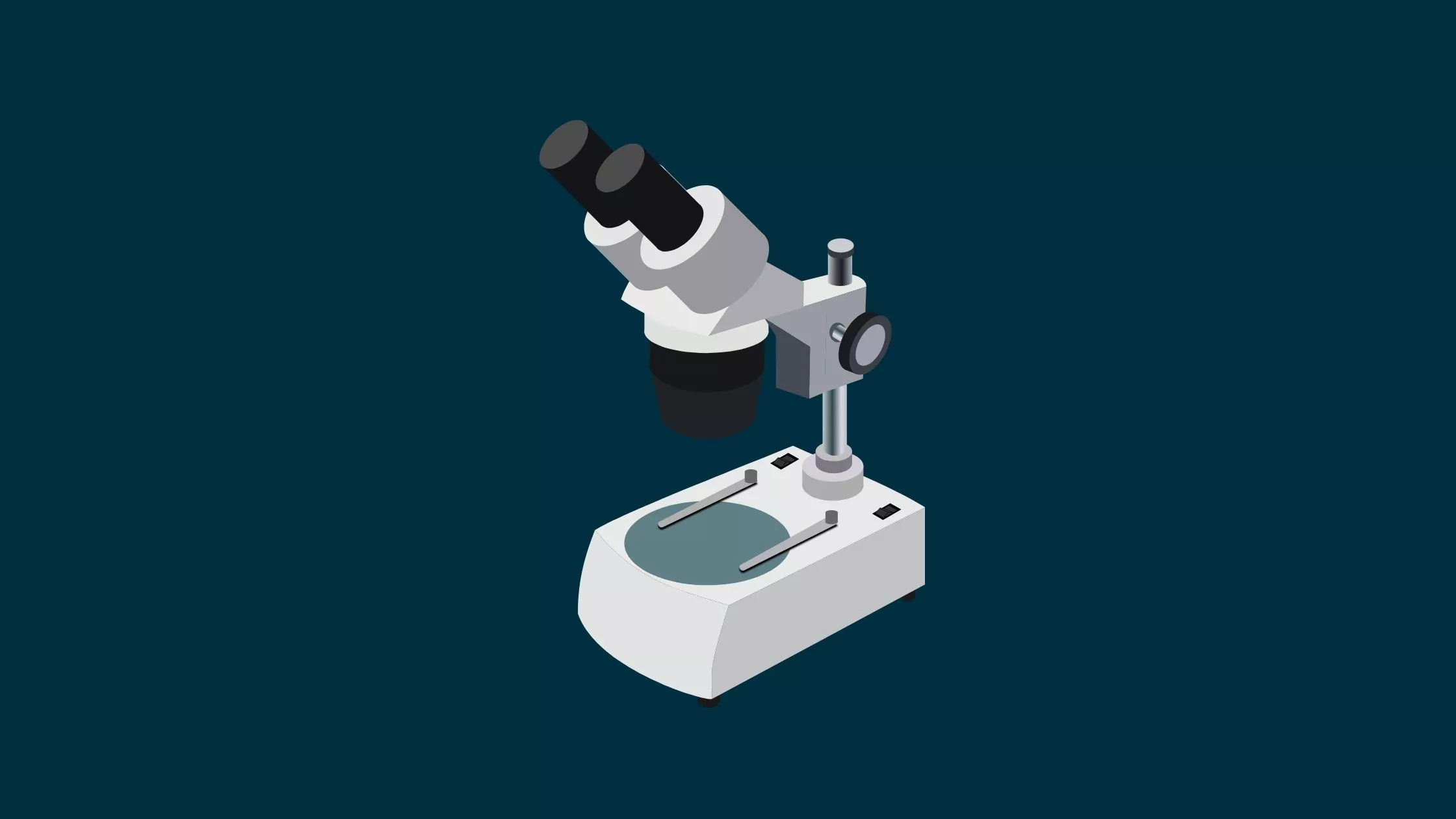
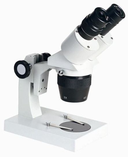
Educational Dual Magnification Stereo Microscope with Built ...
Microscope: Parts Of A Microscope With Functions And Labeled Diagram. Figure: A diagram of a microscope's components The microscope has three basic components: the head, the base, and the arm. Head:Occasionally, the head is considered the body. It holds the optical components of the upper part of the microscope. Base:The microscope's base provides great support. It is also equipped with miniature illuminators.

Compound Microscope Parts, Functions, and Labeled Diagram ...
Compound Microscope Parts, Functions, and Labeled Diagram Eyepiece (ocular lens) with or without Pointer: The part that is looked through at the top of the compound microscope. Eyepieces typically have a magnification between 5x & 30x. Monocular or Binocular Head: Structural support that holds & connects the eyepieces to the objective lenses. Arm: Supports the microscope head and attaches it to the base.
Drawing microscope - Teaching resources
Binocular Microscope Anatomy - Parts and Functions with a Labeled Diagram Let's see the microscope labeled diagram; you will find the flat platform where the slide is placed. Again, this microscope stage lies perpendicular to the optical system or pathway. In some microscopes, the stage can move when the focus is adjusted. Again, the microscope stage is often designed with mechanical devices for holding and moving ...

Compound Microscope Parts, Functions, and Labeled Diagram ...
Light Microscope Labeled Diagram, Definition, Principle, Types, Parts ... Light Microscope Labeled Diagram - Principle of Brightfield Microscope Parts of a bright-field microscope or Compound light microscope An optical microscope, the bright-field microscope (or compound light microscope) is an invaluable tool in the fields of biology, medicine, and education.

A labeled diagram of a microscope. MLT 101. :) | Teaching ...
Microscope, Microscope Parts, Labeled Diagram, and Functions The description given below summarize the brief description of microscope parts used to visualize the microscopic specimens such as animal cells, plant cells, microbes, bacteria, viruses, microorganisms etc. The Microscopes parts divided into three different structural parts Head, Base, and Arms. Head/Body: It contain the optical parts in the ...

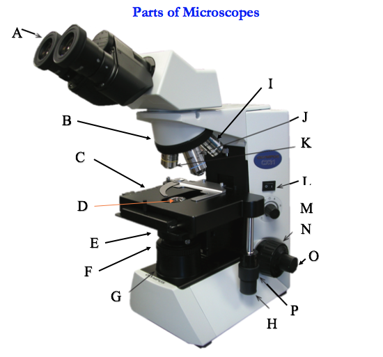
This is a common compound microscope. Label its parts from A ...
16 Essential Microscope Parts: Names, Functions & Labeled Diagram Microscope Parts Labeled Diagram The principle of the Microscope gives you an exact reason to use it. It works on the 3 principles. Magnification Resolving Power Numerical Aperture. Parts of Microscope Head Base Arm Eyepiece Lens Eyepiece Tube Objective Lenses Nose Piece Adjustment Knobs Stage Aperture Microscopic Illuminator Condenser Lens
Light Microscope Labeled Diagram, Definition, Principle ...
Labeled Microscope and Basics of Life Diagram | Quizlet Created by. Chevron_Fickel. A microscope is an instrument widely to magnify and resolve the image of an object that is otherwise invisible to naked eye. For resolving the details of objects, which otherwise cannot be achieved by naked eye, a microscope is used. This set of flash cards will help the student to identify the different parts and ...
Simple Microscope - Parts, Functions, Diagram and Labelling ...
Microscope Parts and Functions Most specimens are mounted on slides, flat rectangles of thin glass. The specimen is placed on the glass and a cover slip is placed over the specimen. This allows the slide to be easily inserted or removed from the microscope. It also allows the specimen to be labeled, transported, and stored without damage.
Labeled Microscope Diagram - Tim's Printables

Compound Microscope Principle, Parts, Diagram Definition ...

Light Microscope- Definition, Principle, Types, Parts ...
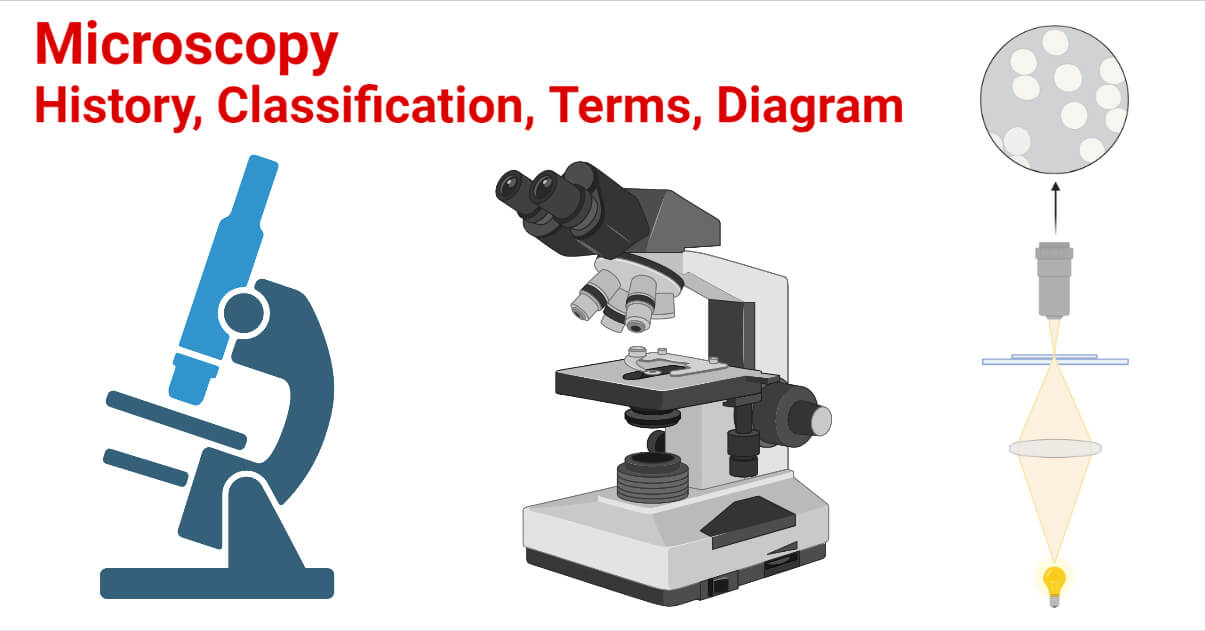
Microscopy- History, Classification, Terms, Diagram

Microscope labeled diagram

Microscope labeled diagram

Compound Microscope – Diagram (Parts labelled), Principle and ...

Parts of a Microscope with Their Functions • Microbe Online

Parts of a Microscope - SmartSchool Systems

Label The Microscope Parts! Diagram | Quizlet

Label the diagram of the microscope and explain the role of ...
Microscope Labeling Game

Diagram of a Microscope by ScienceDoodles on DeviantArt
Addgene: Using a Light Microscope Protocol

Label the microscope — Science Learning Hub

Parts of a microscope with functions and labeled diagram
Parts of a microscope with functions and labeled diagram ...
Instruments of Microscopy | Microbiology | | Course Hero

Microscope diagram labeled | Clipart Panda - Free Clipart Images
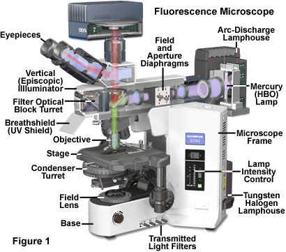
Fluorescence Microscopy - Anatomy of the Fluorescence ...

Guide to understand microscope parts, names, functions & diagram
Experiment 1B | Lab01 | Virtual Edge| General Microbiology ...
Microscope labeled diagram

Microscope Labeling Diagram | Quizlet

Microscope Diagram Labeled Parts - ClipArt Best - ClipArt ...

Microscope With Labels Clip Art at Clker.com - vector clip ...
Labeled Microscope Storyboard by oliversmith
Figure 1.15 A labelled diagram of a light microscope | Boost

Living Environment Course

Label Microscope Diagram - EnchantedLearning.com

Parts of the Microscope with Labeling (also Free Printouts ...
Post a Comment for "45 diagram of a microscope labeled"